An Improved Active Shape Model Based Aortic Segmentation Method for CT Images

An active shape model and CT image technology, applied in the field of medical image processing, can solve the problems of difficult segmentation and extraction of aortic regions, a lot of clinical experience, difficult rescue and other problems, and achieve accurate and reliable experimental results, good adaptability and robustness. Effect

Active Publication Date: 2022-03-04
TIANJIN POLYTECHNIC UNIV
View PDF3 Cites 0 Cited by
  • Summary
  • Abstract
  • Description
  • Claims
  • Application Information

AI Technical Summary

Problems solved by technology

[0002] Aortic dissection is a relatively dangerous type of cardiovascular disease at present. The main reason is the rupture of the aortic wall intima caused by high blood pressure. The wall is ruptured, and rescue is very difficult, and the risk is much higher than that of high-risk conditions such as cerebral infarction, myocardial infarction, and malignant tumors; currently, computerized tomography (Computed Tomography, CT) has become the most important diagnostic method in aortic dissection surgery, but due to every A patient with aortic dissection has many CT scan images, usually 500 to 1000. Generally, cardiologists need to analyze the patient’s CT scan image information one by one. The optimal timing of treatment is missed; therefore, domestic and foreign medical diagnostic equipment developers usually use CT scan image sequences of the thoracoabdominal cavity of patients with aortic dissection, and use image processing methods to perform three-dimensional reconstruction of the outer contour of the aorta to assist physicians in understanding the three-dimensional structure of the patient's aorta. Have a relatively three-dimensional and comprehensive understanding of the location and size of the hematoma in detail. The most important thing in the reconstruction process is to accurately segment the aortic region; because the location and shape of the aorta vary greatly among different individuals, and there are other The interference of organs and tissues makes it difficult to accurately segment and extract the aortic region in CT images; currently, the segmentation algorithms for medical CT images mainly include methods based on regions, boundaries and shape models; the main advantages of region-based segmentation methods It is simple and efficient, but it is difficult to obtain satisfactory segmentation results for images with insignificant grayscale differences between different target regions; the boundary-based method is better for image segmentation with obvious edges and low noise, but it is sensitive to noise. Images with complex edges are prone to false edges or discontinuous edges; the advantage of the algorithm based on the active shape model is that it does not need to select the region of interest, and can directly input the entire CT image to achieve segmentation and extraction, which is widely used in practical applications, but when the target segmentation When there is a large difference between some regions and the trained aorta model, there is a certain error; therefore, it is difficult to develop an automatic and accurate extraction algorithm for various aortic region structures in human thoracoabdominal CT images

Method used


Image

  • An Improved Active Shape Model Based Aortic Segmentation Method for CT Images
  • An Improved Active Shape Model Based Aortic Segmentation Method for CT Images
  • An Improved Active Shape Model Based Aortic Segmentation Method for CT Images

Examples


Embodiment Construction

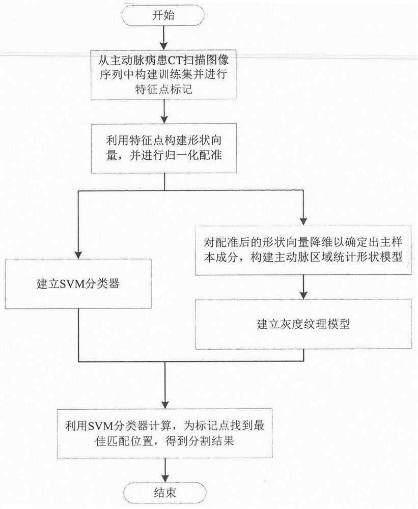
[0024] Algorithm flow chart of the present invention is as figure 1 As shown, first select some samples from the CT scan image sequence of aortic patients to build a training set, and mark the aortic region in the sample with feature points; the marked points of each CT image constitute a shape vector, and then all the constructed The shape vector normalization registration of the shape vector; in order to simplify the calculation of high-dimensional data, the principal component analysis method is used to reduce the dimension of the registered data to determine the main sample components, and build a statistical shape model; then the data after dimension reduction is established Grayscale texture model; establish SVM classifier in the training process; use SVM classifier to calculate target contour probability P(i, j) in the set of marker points, allowing the evolution of the outline to be based on the model outline. The combination of optimization and target contour and mode...


PUM


Abstract

The invention discloses an improved CT image aorta segmentation method based on an active shape model, which adopts a segmentation extraction method combining a support vector machine and an active shape model. The precise segmentation and extraction of the region solves the problem of errors caused by the large difference between the model and the actual segmentation target in the existing algorithm; the process is as follows: (1) select samples from the CT images of aortic patients to construct a training set, and the main The arterial area is marked with feature points; (2) the feature points of the sample mark are constructed into a shape vector and normalized for registration; (3) the dimensionality of the vector is reduced to determine the main sample components, and a statistical shape model is constructed; (4) the feature points Points as the center of the square matrix for grayscale sampling to establish a texture model; (5) build a support vector machine classifier during model training; (6) calculate the probability of the marked point set to the target contour, and find the best matching position.

Description

technical field [0001] The invention belongs to the technical field of medical image processing and relates to an improved active shape model-based CT image aorta segmentation and extraction method, which can be used for automatic segmentation and extraction of the aorta region in the CT scan image of the human chest and abdominal cavity. Background technique [0002] Aortic dissection is a relatively dangerous type of cardiovascular disease at present. The main reason is the rupture of the aortic wall intima caused by high blood pressure. The wall is ruptured, and rescue is very difficult, and the risk is much higher than that of high-risk conditions such as cerebral infarction, myocardial infarction, and malignant tumors; currently, computerized tomography (Computed Tomography, CT) has become the most important diagnostic method in aortic dissection surgery, but due to every A patient with aortic dissection has many CT scan images, usually 500 to 1000. Generally, cardiolog...

Claims


Application Information

Patent Timeline
Patent Type & Authority Patents(China)
IPC IPC(8): G06T7/11G06T7/33G06V10/764G06V10/77
CPCG06T7/11G06T7/33G06T2207/30101G06T2207/10081G06F18/2135G06F18/2411
Inventor 段晓杰左瑞雪汪剑鸣张美松石小兵王琦李秀艳
Owner TIANJIN POLYTECHNIC UNIV
Why Eureka
Try Eureka