
Fluorescent biosensor based on cyclic amplification technology and carboxyl carbon quantum dots as well as preparation method and application of fluorescent biosensor

A technology of biosensors and carbon quantum dots, applied in the field of biosensors, can solve the problems of complex and strict preparation of nanomaterials, difficulty in ensuring the biocompatibility of nanomaterials, etc., and achieve excellent accuracy, great application potential, and rapid analysis and detection Effect

Inactive Publication Date: 2019-03-08
QINGDAO UNIV OF SCI & TECH
View PDF6 Cites 3 Cited by
  • Summary
  • Abstract
  • Description
  • Claims
  • Application Information

AI Technical Summary

Problems solved by technology

Although these nanomaterials have been successfully used as nano-quenchers for the detection of nucleic acids, the preparation of some of them is often complicated and rigorous, and it is difficult to ensure that some of them have good biocompatibility [Liao K H, Lin Y S, Macosko C W, et al. ACS applied materials & interfaces, 2011, 3(7): 2607-2615.]

Method used


Image

  • Fluorescent biosensor based on cyclic amplification technology and carboxyl carbon quantum dots as well as preparation method and application of fluorescent biosensor
  • Fluorescent biosensor based on cyclic amplification technology and carboxyl carbon quantum dots as well as preparation method and application of fluorescent biosensor
  • Fluorescent biosensor based on cyclic amplification technology and carboxyl carbon quantum dots as well as preparation method and application of fluorescent biosensor

Examples


Embodiment 1

[0021] Example 1. Preparation of fluorescent biosensor and detection of miR34c

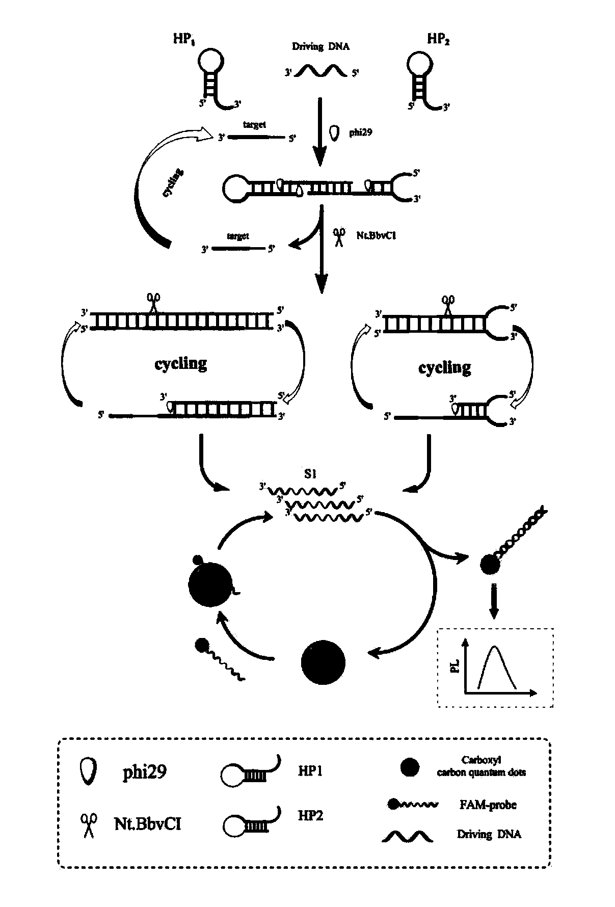
[0022] Target-assisted cyclic amplification process: Before the hairpin structure is used, hairpin 1 (HP1) and hairpin 2 (HP2) are heated in a water bath at 90 °C for 5 min to form a hairpin structure, and then slowly cooled to room temperature to form stem-loop DNA structure. Take 6 μL Tris buffer, 2 μL HP1 (10 μM), 2 μL HP2 (10 μM), 2 μL DP1 (10 μM) and 2 μL target (MicroRMA-34) (different concentrations), respectively, add 2 μL 10×phi29 polymerase Buffer, 2 μL 10× NEB Cutsmart Buffer, 0.5 μL phi29 (10,000 U / ml) polymerase, 0.5 μL Nt.BbvCI (10 U / μL) nickase and 1 μL dNTPs (10 mM) constitute a 20 μL cyclic amplification reaction system. The above reaction system was placed in a constant temperature oscillator at 37°C to react for 3.5h. Finally, the reaction was terminated by placing at 80 °C for 20 min to obtain S1.

[0023] Fluorescence quenching process: In order to achieve fluorescence quen...

Embodiment 2

[0025] Example 2. Preparation of fluorescent biosensor and detection of miR34c

[0026] Changed "First add 40 μL cCQD to 40nM FAM-probe fluorescent probe, incubate at 50°C for 30 min." to "First add 30 μL cCQD to 40nM FAM-probe fluorescent probe, incubate at 50°C 30min." Other preparation conditions were the same as in Example 1, and a biosensor with morphology and properties similar to those in Example 1 was obtained. The results of the detection of miR34c were the same as those in Example 1.

Embodiment 3

[0027] Example 3. Preparation of fluorescent biosensor and detection of miR34c

[0028] Will "take 6μL Tris buffer, 2μL HP1 (10μM), 2μL HP2 (10μM), 2μL DP1 (10μM) and 2μL target (MicroRMA-34) (different concentrations), add 2μL 10 × phi29 polymerase Buffer, 2μL 10×NEBCutsmart Buffer, 0.5μL phi29 (10,000U / ml) polymerase, 0.5μL Nt.BbvCI (10U / μL) nickase and 1μL dNTPs (10mM) constitute a 20μL cyclic amplification reaction system.” To "take 6 μL Tris buffer, 2 μL HP1 (10 μM), 2 μL HP2 (10 μM), 2 μL DP1 (10 μM), and 2 μL target (MicroRMA-34) (different concentrations), respectively, add 2 μL 10×phi29 polymerase Buffer, 2 μL 10×NEB Cutsmart Buffer, 0.4μL phi29 (10,000U / ml) polymerase, 0.4μL Nt.BbvCI (10U / μL) nickase and 1μL dNTPs (10mM) constitute a 20μL cyclic amplification reaction system.” Other preparation conditions were the same as those in Example 1, and a biosensor with morphology and properties similar to those in Example 1 was obtained. The results of the detection of mi...


PUM


Abstract

The invention discloses a fluorescent biosensor based on an enzyme-catalyzed target cyclic amplification technology combined with carboxyl functionalized carbon quantum dots as well as a preparation method and application of the fluorescent biosensor. A technical solution adopted in the invention is that a tumor-associated gene miRNA34c is used as a fuel molecule to design a multi-wheel driven DNAnanomachine comprising an unmarked stem-loop structure 1 (HP1), a stem-loop structure 2 (HP2) and a driving primer (DP) for cyclic amplification of a trace target. In addition, the synthesized carboxyl carbon quantum dots (cCQD) with excellent biocompatibility firstly is bound to a fluorescent probe to quench a fluorophore; an S1 cycle product in a target stimulating cyclic amplification processis added and placed for 20 min at room temperature to recover fluorescence; then, a fluorescence intensity of the mixture is measured so as to realize sensitive detection of the target miRNA34c. The research idea provides a new strategy for the sensitive detection of miRNA34c and is expected to be used in early diagnosis of tumors.

Description

Technical field: [0001] The invention relates to a biosensor based on the enzymatic target cyclic amplification technology combined with carboxyl-functionalized carbon quantum dot fluorescent probes; the invention also relates to a preparation method of the biosensor and its analytical application for detecting miRNA. Background technique: [0002] MicroRNAs (miRNAs) are a class of endogenous non-coding RNA molecules (20-23nt) that are targeted for degradation by translational inhibition or by forming RNA-induced silencing complex (RISC) with target messenger RNAs. Physiological processes such as proliferation, development, metabolism, immune response, tumor, and viral infection in biology have a crucial impact [Lee I, Ajay S S, Chen H, et al. Nucleic Acids Research, 2008, 36(5): e27.]. Due to the low abundance and easy degradation of miRNAs, direct detection can only be detected at trace amounts. Therefore, there is an urgent need to develop sensitive, highly selective, co...

Claims


Application Information

Patent Timeline
Patent Type & Authority Applications(China)
IPC IPC(8): G01N21/64C12Q1/6825
CPCG01N21/6428G01N21/6486C12Q1/6825G01N2021/6432
Inventor 接贵芬李春丽
Owner QINGDAO UNIV OF SCI & TECH
Learn More