Method for separating exosomes by low-speed ultrafiltration centrifugation conjugated polymer precipitation technique

A technology combining polymers and exosomes, applied in the field of biomedicine, can solve the problems of large amount of reagents used, inability to purchase polymer precipitation method kits in large quantities, high price, etc., achieving simple and easy operation methods and overcoming background impurities. The effect of interference and reducing the cost of reagent use

Inactive Publication Date: 2019-04-12
JINAN UNIVERSITY
View PDF7 Cites 8 Cited by
  • Summary
  • Abstract
  • Description
  • Claims
  • Application Information

AI Technical Summary

Problems solved by technology

Ultra-high-speed centrifugation requires special and expensive instruments. At present, the purchase cost of an ultra-high-speed centrifuge alone is more than 100,000 yuan, and the separation is often due to protein contamination and exosome rupture. Universal application
The simple polymer precipitation method has already launched products, but the use of reagents is large and the price is expensive, which affects further experiments.
Moreover, due to the decreased division ability of nerve cells, the amount of exosomes secreted by tumor cells is less, making separation and enrichment more difficult.
In order to overcome the lack of ultra-high-speed centrifuges in most existing laboratories, only conventional speed centrifuges, and the lack of funds to purchase a large number of polymer precipitation kits, etc., the development of isolated exosomes has strong operability, high purity, and Affordable methods are especially urgently needed

Method used


Image

  • Method for separating exosomes by low-speed ultrafiltration centrifugation conjugated polymer precipitation technique
  • Method for separating exosomes by low-speed ultrafiltration centrifugation conjugated polymer precipitation technique
  • Method for separating exosomes by low-speed ultrafiltration centrifugation conjugated polymer precipitation technique

Examples


Embodiment 1

[0042] Example 1 Extraction and purification of nerve cell exosomes

[0043]1. Extract primary neurons (including cortical neurons and hippocampal neurons) from newborn mice: use ethanol to sterilize the head of newborn mice within 24 hours (balb / c mice were purchased from Guangzhou University of Traditional Chinese Medicine), cryoanesthetize, and place in The cortex and hippocampus were separated by PBS phosphate buffered saline at 4°C, and the meninges and blood vessels were removed. After resting for 2 minutes, the supernatant was discarded, 0.125% trypsin (Gibico Company) was added to the tissue, the digestion was terminated after 10 minutes of digestion, the cells were resuspended, and the supernatant was discarded after centrifugation at 1000 r / min for 5 minutes, and plated. The complete medium DMEM / F12 (purchased from Gibico Company) was used to promote neuron maturation, and the serum-free medium containing nerve cell growth factor (Neurobasal+2% B27 medium from Gibico...

Embodiment 2

[0051] Example 2 Identification of exosomes

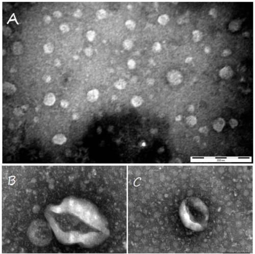
[0052] 1. Morphological observation

[0053] The morphology of the exosomes obtained in Example 1 was observed by a transmission electron microscope.

[0054] (1) After fully mixing the exosomes obtained in Example 1 with PBS;

[0055] (2) Add 20 μL to the sample-loading copper grid with a diameter of 2 mm, and let it stand for 5 minutes;

[0056] (3) Use filter paper to gently blot the liquid around the copper mesh, then place the copper mesh upside down on a 20g / L phosphotungstic acid (pH6.8) drop, and negatively stain at room temperature for 10 minutes;

[0057] (4) The copper grid was dried under an incandescent lamp, and placed under a transmission electron microscope to find a vesicle-like structure with a diameter of 40-100 nm and take pictures. The results are as follows: figure 1 As shown, the scale of A is 200nm, B and C are enlarged compared with A, and the scales are 50nm and 100nm respectively.

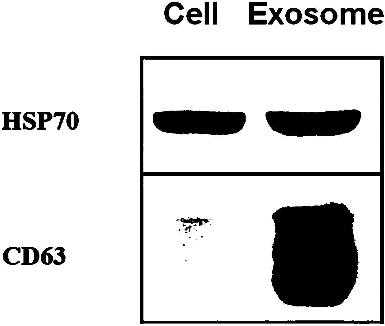
[0058] 2. Labeled ...

Embodiment 3

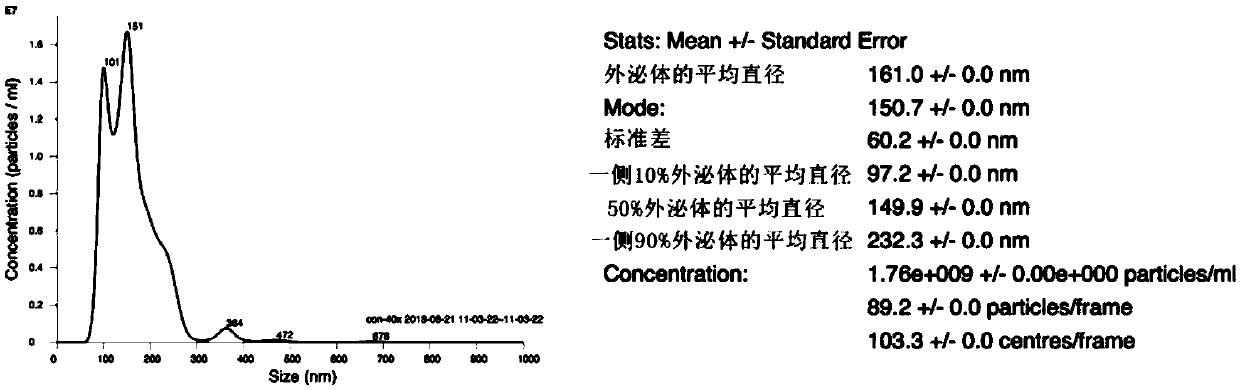
[0075] Example 3 Study on particle diameter and concentration of exosomes

[0076] The nanoparticle tracer technology further identifies exosomes, and the concentration and particle diameter of exosomes are obtained by analysis, detection and purification. Nanosight visual nanoparticle analyzer can directly and dynamically observe the state of Brownian motion of nanoscale particles in the sample, and use the Stroke-Einstein equation to directly obtain the particle size of each particle, and can measure the true concentration of particles (units / ml). The result is as image 3 As shown, the concentration, size and other characteristics of the product obtained in the present invention are in line with the expression of exosomes.


PUM

PropertyMeasurementUnit
diameteraaaaaaaaaa

Abstract

The invention provides a method for separating exosomes by a low-speed ultrafiltration centrifugation conjugated polymer precipitation technique. The method comprises the steps of performing concentration through a low-speed centrifugation and ultrafiltration centrifugal tube, and through combination with a polymer precipitation technique, separating the exosomes, wherein only a tabletop centrifuge common in laboratory is needed, and only the ultrafiltration centrifugal tube low in cost is needed. The operation method is simple and easy to operate, and the bioactivity of the exosomes is not influenced. The method also effectively solves the problems that compared with other cells, nerve cells are low in the quantity of secretion exosomes and difficult to separate and enrich. Besides, liquid containing the exosomes is effectively concentrated, and a polymer reagent which is used is greatly reduced. Compared with the prior art, the separation purity and the efficiency are higher, the maneuverability is higher, the content of the separated exosomes can be reduced, and the method has a favorable scientific application promotion value and favorable application prospects.

Description

technical field [0001] The invention belongs to the technical field of biomedicine and relates to a method for separating exosomes by combining a low-speed centrifuge and an ultrafiltration centrifuge tube, in particular to a method for separating exosomes by combining low-speed ultrafiltration centrifugation with polymer precipitation technology. Background technique [0002] As a kind of microvesicles with a diameter of 30-100nm that are actively secreted by cells, exosomes are important mediators of intercellular communication and participate in diseases including cancer, infectious diseases and neurodegenerative diseases. Signal transmission between biological cells. Clinically, exosomes can serve as biomarkers, drug carriers, explain the mechanism of disease occurrence, and display new targets for intervention. Exosomes secreted by nerve cells are the carrier of communication between nerve cells, and play an important regulatory role in the occurrence, development and ...

Claims


Application Information

Patent Timeline
Patent Type & Authority Applications(China)
IPC IPC(8): C12N5/079
CPCC12N5/0618C12N2501/13C12N2509/00
Inventor 逯丹麦鸿成臧健坤吴有盛徐安定
Owner JINAN UNIVERSITY
Why Eureka
Try Eureka