A peanut chromosome probe staining kit and its application method
A dyeing reagent and chromosome technology, which is applied in the field of peanut cytology research, can solve the problems of multi-manual operation steps, unfavorable automatic identification, etc., and achieve the effect of simple technical method and high cost
- Summary
- Abstract
- Description
- Claims
- Application Information
AI Technical Summary
Problems solved by technology
Method used
Image
Examples
Embodiment 1
[0026] Embodiment 1, peanut chromosome probe staining kit
[0027] The kit includes Probe Mix dry powder, DAPI stock solution with a concentration of 100 μg / ml, 2×SSC buffer solution, DAPI buffer solution, staining jar and variety control karyotype.
[0028] The probes in Probe Mix dry powder include TAM fluorescently labeled probes DP-1, DP-7 and FAM fluorescently labeled probes DP-5, DP-8, each probe content is 0.2OD, as follows:
[0029]
[0030] 2 x SSC buffer consisting of 0.3 M trisodium citrate C 6 h 5 Na 3 o 7 2H 2 O and 3M NaCl composition.
[0031] DAPI buffer consists of 0.1M citric acid and 0.05M Na 2 HPO 4 composition.
[0032] The staining jar is a black light-proof glass staining jar.
Embodiment 2
[0033] Embodiment 2, the use method of peanut chromosome probe staining kit, comprises the following steps:
[0034] (1) Preparation of dyeing solution: Dissolve Probe Mix dry powder in 1ml 2×SSC buffer, pour it into the staining jar, and add 39ml 2×SSC buffer to make the final concentration of DP-1, DP-5, DP-7 and DP-8 Both are 0.005OD / ml;
[0035] (2) Put the slides (76×26×1mm) containing the peanut chromosomes in the staining solution of the staining jar, and stain at 35-37°C for 6 hours; take out the slides, wash them with 2×SSC buffer for 3 times, and then Add 500 μl of the mixture of DAPI mother solution (100 μg / ml) and DAPI buffer solution with a volume ratio of 2.5:500 dropwise on each slide, stain for 3-4 minutes, then wash with DAPI buffer solution for 4-5 times, and drop 5 μl of antibacterial solution. Fluorescence-quenched mounting medium, covered with a coverslip, and photographed under a fluorescent microscope.
Embodiment 3
[0036] Embodiment 3, the staining of the four red chromosomes of peanut cultivars
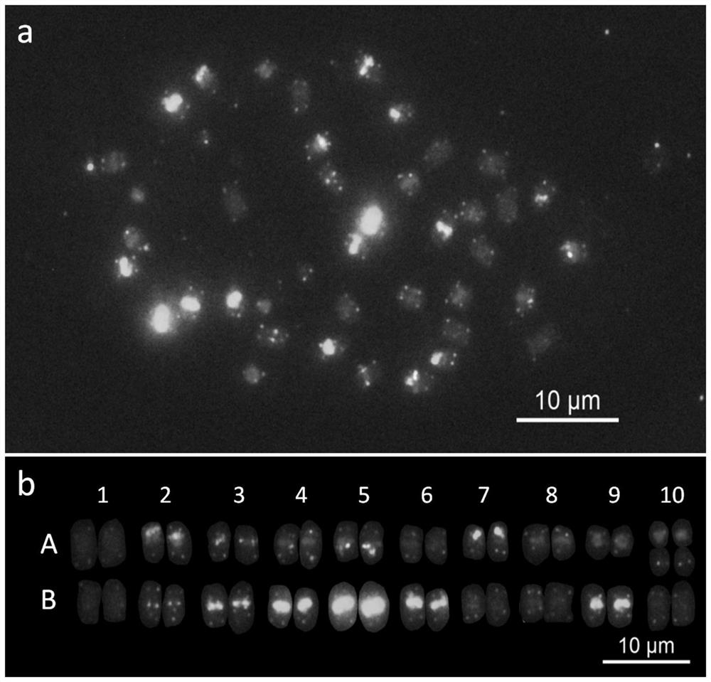
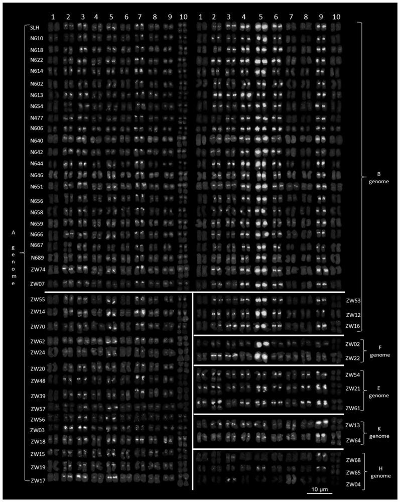
[0037] Adopt the method for embodiment 2, use the same dyeing vat dyeing solution to dye the peanut cultivar four-grain red metaphase chromosome, and dye to obtain the picture ( figure 1 a), and cut out each chromosome, and arrange the karyotype ( figure 1 b). from figure 1 It can be seen from the figure that after staining with the Probe Mix dye solution of the kit, there are obvious red and green characteristic signals on the chromosomes, the A genome is dominated by red signals, and the B genome is dominated by green signals, and A and B of peanuts can be clearly distinguished according to the color Genome. In addition, the banding patterns of peanut chromosomes are different, and the characteristics are as follows: In genome A, there is only a weak signal at the end of chromosome 1, weaker red and green signals at the centromere of chromosome 2, and weaker red and green signals at the ce...
PUM
| Property | Measurement | Unit |
|---|---|---|
| concentration | aaaaa | aaaaa |
Abstract
Description
Claims
Application Information
 - R&D
- Intellectual Property
- Life Sciences
- Materials
- Tech Scout
- Unparalleled Data Quality
- Higher Quality Content
- 60% Fewer Hallucinations
Browse by: Latest US Patents, China's latest patents, Technical Efficacy Thesaurus, Application Domain, Technology Topic, Popular Technical Reports.
© 2025 PatSnap. All rights reserved.Legal|Privacy policy|Modern Slavery Act Transparency Statement|Sitemap|About US| Contact US: help@patsnap.com


