Fluorescent microsphere combined detection device for cardiac marker and preparation method of fluorescent microsphere combined detection device

A technology of fluorescent microspheres and combined detection, which is applied in the medical field, can solve the problems of easy quenching of fluorescent antibodies, insufficient amount of adsorbed protein on NC membrane, weak binding force, etc., achieve good biocompatibility, and improve coating efficiency , the effect of high mechanical strength

Inactive Publication Date: 2019-05-24
JILIN UNIV +1
View PDF9 Cites 9 Cited by
  • Summary
  • Abstract
  • Description
  • Claims
  • Application Information

AI Technical Summary

Problems solved by technology

[0010] The present invention provides a fluorescent microsphere joint detection device for cardiac markers and a preparation method thereof, so as to solve the problems in the prior art that the fluorescent antibody is easily quenched, the amount of protein adsorbed by the NC membrane is insufficient, and the binding force is not strong

Method used


Image

  • Fluorescent microsphere combined detection device for cardiac marker and preparation method of fluorescent microsphere combined detection device
  • Fluorescent microsphere combined detection device for cardiac marker and preparation method of fluorescent microsphere combined detection device
  • Fluorescent microsphere combined detection device for cardiac marker and preparation method of fluorescent microsphere combined detection device

Examples


Embodiment 1

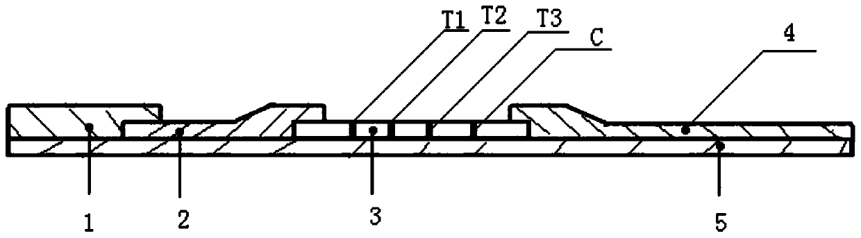
[0043] Example 1 A fluorescent microsphere combined detection device for cardiac markers

[0044] The sample pad 1, the immunofluorescence antibody glass fiber membrane 2, the nitrocellulose membrane 3, and the absorption pad 4 are pasted on the plastic plate 5 respectively, and the two ends of the nitrocellulose membrane 3 are connected with the absorption pad 4, the immunofluorescence antibody glass fiber respectively. The membrane 2 is overlapped, and the other end of the immunofluorescence antibody glass fiber membrane 2 is overlapped with the sample pad 1; the nitrocellulose membrane 3 is provided with a first detection line T1, a second detection line T2, and a third detection line T3 , and quality control line C; the solid phase of the first detection line T1 has a highly specific MPO antibody, the solid phase of the second detection line T2 has a high specificity cTnI antibody, and the third detection line There is a highly specific NT-proBNP antibody on the solid phas...

Embodiment 2

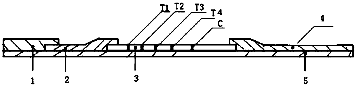
[0045] Example 2 A fluorescent microsphere combined detection device for cardiac markers

[0046] On the basis of Example 1, a fourth detection line T4 is further set on the nitrocellulose membrane 3, and the solid phase of the fourth detection line T4 has a highly specific D-dimer antibody.

Embodiment 3

[0048] The preparation method of the fluorescent microsphere combined detection device for cardiac markers in the above-mentioned embodiment 1

[0049] Include the following steps:

[0050](a) Preparation of immunofluorescent microspheres, take 100ul of microsphere suspension with a solid content of 1%, dilute it 10 times with ultrapure water, that is, 1000ul, add it to a test tube, and take 40ul of N-hydroxysuccinimide solution Add (NHS) to the microsphere suspension and mix well, then add 40ul of 1-(3-dimethylaminopropyl)-3-ethylcarbodiimide hydrochloride (EDC) to the microsphere suspension Mix well in the medium, and react at room temperature for 0.5 hours; ultrasonically sonicate the reacted suspension to resuspend the microspheres on the tube wall in the aqueous solution, then centrifuge the microsphere suspension at 8000r / min, 15min, pour Remove the supernatant, add 1ml ultrapure water, and then disperse evenly with ultrasonic waves;

[0051] (b) Immunofluorescent micr...


PUM


Abstract

The invention relates to a fluorescent microsphere combined detection device for a cardiac marker and a preparation method of the fluorescent microsphere combined detection device, and belongs to thefield of medical detection equipment. A sample pad, an immunofluorescent antibody glass fiber membrane, a nitrocellulose membrane and an absorption pad respectively adhere to a plastic plate, the twoends of the nitrocellulose membrane are respectively overlapped with the absorption pad and the immunofluorescent antibody glass fiber membrane, the other end of the immunofluorescent antibody glass fiber membrane is overlapped with the sample pad, the nitrocellulose membrane is provided with a first detection line T1 with a solid-phase high-specificity MPO antibody, a second detection line T2 with a solid-phase high-specificity cTnI antibody, a third detection line T3 with a solid-phase high-specificity NT-proBNP antibody, a fourth detection line T4 with a solid-phase high-specificity D dimerantibody and a quality control line C; and a goat anti-mouse IgG polyclonal antibody is sprayed on the quality control line C. The fluorescent microsphere combined detection device for the cardiac marker has the advantages that the sensitivity and the specificity of reaction are effectively improved, the use amount of immune microspheres can further be reduced under the same threshold value, thecost is saved, and the device is simple and convenient to operate and high in practicability.

Description

technical field [0001] The present invention relates to the field of medical detection equipment, in particular to a combined detection device for cardiac markers and a preparation method thereof, which uses fluorescence immunochromatography technology and the principle of double-antibody sandwich method to quantitatively detect human myeloperoxidase (MPO) in plasma of clinical specimens. ), cardiac troponin I (cTnI), N-terminal pro-brain natriuretic peptide (NT-proBNP) and D-dimer detection device and preparation method thereof, which can realize sensitive, specific and rapid detection of cardiac markers. Background technique [0002] Cardiopulmonary function indicators are an important reference for clinical diagnosis and disease prevention. Monitoring cardiopulmonary function is of great value in the diagnosis, treatment and prognosis of patients with chronic cardiopulmonary diseases. The detection of cardiopulmonary function indicators is widely used in clinical emergenc...

Claims


Application Information

Patent Timeline
Patent Type & Authority Applications(China)
IPC IPC(8): G01N33/533G01N33/68
Inventor 王珺楠杨小军李欣赵旻
Owner JILIN UNIV
Why Eureka
Try Eureka