Tendon-bone combined three-phase scaffold prepared by fusion electrospinning three-dimensional printing

A technology of 3D printing and fused static electricity, which is applied in the medical field, can solve problems such as easy tissue fracture, failure to restore gradient structure, and inability of fibrous scar tissue to effectively disperse stress, so as to promote cell proliferation and repair, facilitate tendon-bone joint repair and Restoration of normal function, high precision effect

Inactive Publication Date: 2019-06-04
SHANGHAI NINTH PEOPLES HOSPITAL AFFILIATED TO SHANGHAI JIAO TONG UNIV SCHOOL OF MEDICINE
View PDF2 Cites 5 Cited by
  • Summary
  • Abstract
  • Description
  • Claims
  • Application Information

AI Technical Summary

Problems solved by technology

[0003] However, in this treatment method, the tendon-bone junction will be filled with fibrous scar tissue in the early stage, and the gradient structure of bone, cartilage, and tendon in the defect area cannot be restored. The regenerated fibrous scar tissue cannot effectively disperse the stress, and after returning to normal activities, This tissue is easy to break again. Therefore, in treatment, in addition to improving surgical skills and rehabilitation plans, an effective method is needed to completely reconstruct the gradient struct

Method used


Image

  • Tendon-bone combined three-phase scaffold prepared by fusion electrospinning three-dimensional printing
  • Tendon-bone combined three-phase scaffold prepared by fusion electrospinning three-dimensional printing
  • Tendon-bone combined three-phase scaffold prepared by fusion electrospinning three-dimensional printing

Examples


Embodiment 1

[0025] The present invention provides the following technical solutions: tendon-bone joint three-phase scaffold prepared by melt electrospinning three-dimensional printing, the specific examples are as follows:

[0026] Specifically, tendon stem cells are cultured in an incubator with DMEM medium containing 10% fetal bovine serum, chondrocytes are cultured in an incubator with F12 medium containing 10% fetal bovine serum, and bone marrow stem cells are cultured in a medium containing 10% fetal bovine serum. , connective tissue growth factor CTGF 25ng / ml, ascorbic acid 25uM α-MEM medium cultured in the incubator.

[0027] Specifically, a three-dimensional printing software is used to establish a printing model, which is in the shape of a multi-layer rectangle, and the path gaps of the tendon area, cartilage area, and bone area are adjusted and saved according to needs.

[0028] Specifically, appropriate amounts of biopolymer materials A1, A2, and A3, and microspheres B1, B2, an...

Embodiment 2

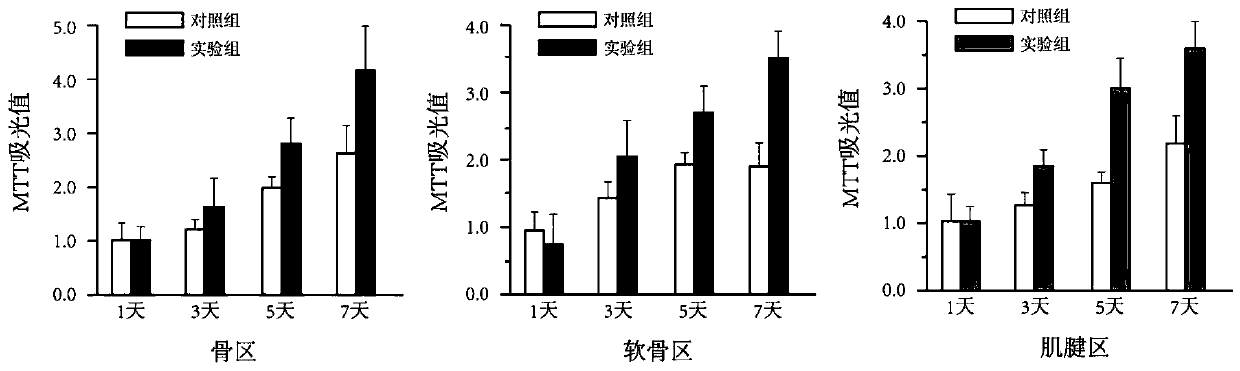
[0031] Corresponding cells were planted in each area of ​​the obtained three-phase integrated scaffold, and cultured in an incubator. Tendon stem cells, chondrocytes, and bone marrow stem cells were implanted in the scaffolds obtained by three-dimensional printing of pure polycaprolactone as the control group, respectively. Each area of ​​the scaffold was compared. After culturing for 1, 3, 5, and 7 days respectively, the tenocyte viability in the tendon scaffold was detected by MTT method. The detection results were as follows: figure 1 As shown, the cells on the scaffold obtained by the melt electrospinning three-dimensional printing technology with cytokine microspheres have good proliferation behavior, and the cell viability is better, and the proliferation behavior is more significant than that of the control group.

Embodiment 3

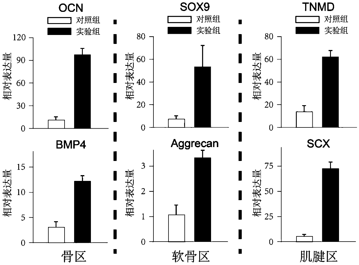
[0033] The obtained three-phase integrated scaffold was planted with corresponding cells in each area, and the qRT-PCR test was performed. The scaffolds obtained by three-dimensional printing of polycaprolactone were respectively planted with tendon stem cells, chondrocytes, and bone marrow stem cells as a control group, and cultured separately. After 7 days, qRT-PCR detection was carried out, and the detection results were as follows: figure 2 As shown, the expression of tendon-related products (SCX, TNMD) in the tendon region, cartilage-related products (SOX9, Aggrecan) in the cartilage region, and bone-related products (OCN, BMP4) in the bone region in the experimental group were significantly higher.


PUM


Abstract

The invention belongs to the technical field of medical treatment, and particularly relates to a tendon-bone combined three-phase scaffold prepared by fusion electrospinning three-dimensional printing. A method comprises the following steps: S1, cell cultivation; S2, bracket preparation; and S3, cell planting. The method can accurately control the fiber diameter and the printing path by using a fusion electrostatic spinning three-dimensional printing technology, thereby greatly improving the printing precision; by using a spinning solution containing cytokine microspheres for electrospinning printing, the electrospinning fibers containing a large amount of cytokines can be obtained; and through different three-dimensional printing path parameters, the tendon-cartilage-bone three-phase scaffold can be obtained by printing. The scaffold obtained by the method has a bone-cartilage-tendon tissue gradient structure, has high structure precision, carries a large amount of microspheres with cytokines for slow release, promotes cell proliferation repair, and is beneficial to tendon-bone combined and normal function recovery.

Description

technical field [0001] The invention belongs to the field of medical technology, and in particular relates to a tendon-bone joint three-phase support prepared by melt electrospinning three-dimensional printing. Background technique [0002] Tendon-bone syndesmosis injury is one of the common sports injuries, and it is also a common type of clinical soft tissue injury. Tendons and ligaments are directly fixed on the bone, and the normal function of the area is restored as much as possible; [0003] However, in this treatment method, the tendon-bone junction will be filled with fibrous scar tissue in the early stage, and the gradient structure of bone, cartilage, and tendon in the defect area cannot be restored. The regenerated fibrous scar tissue cannot effectively disperse the stress, and after returning to normal activities, This tissue is easy to break again. Therefore, in treatment, in addition to improving surgical skills and rehabilitation plans, an effective method is...

Claims


Application Information

Patent Timeline
IPC IPC(8): C12M3/00B33Y30/00
Inventor 乔之光唐佳昕戴尅戎孙彬彬王友连梅菲
Owner SHANGHAI NINTH PEOPLES HOSPITAL AFFILIATED TO SHANGHAI JIAO TONG UNIV SCHOOL OF MEDICINE
Why Eureka
Try Eureka