Nanofiber antitumor vaccine formed by self-assembly peptide folding and method
A technology of nanofibers and self-assembled peptides, applied in the fields of molecular biology and immunology, can solve problems such as limited immunogenicity, inability to produce a strong enough anti-tumor immune response, lack of curative effect of vaccines, etc., achieve significant healing effect, suppress immunity Effect of suppressor cell MDSC, significant anti-tumor immune effect
- Summary
- Abstract
- Description
- Claims
- Application Information
AI Technical Summary
Problems solved by technology
Method used
Image
Examples
Embodiment 1
[0042] Example 1: Preparation and morphological identification of nanofiber anti-tumor vaccine
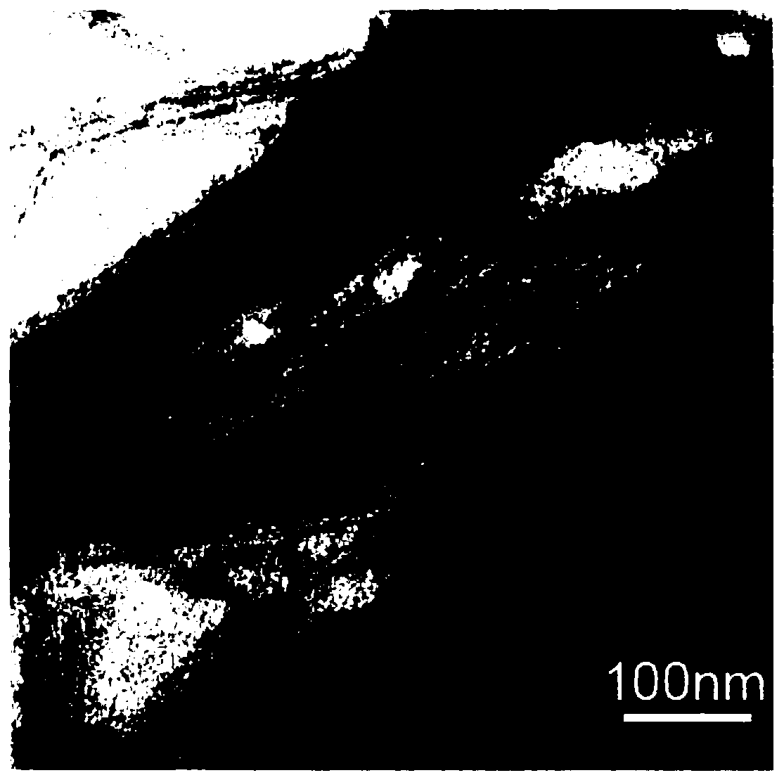
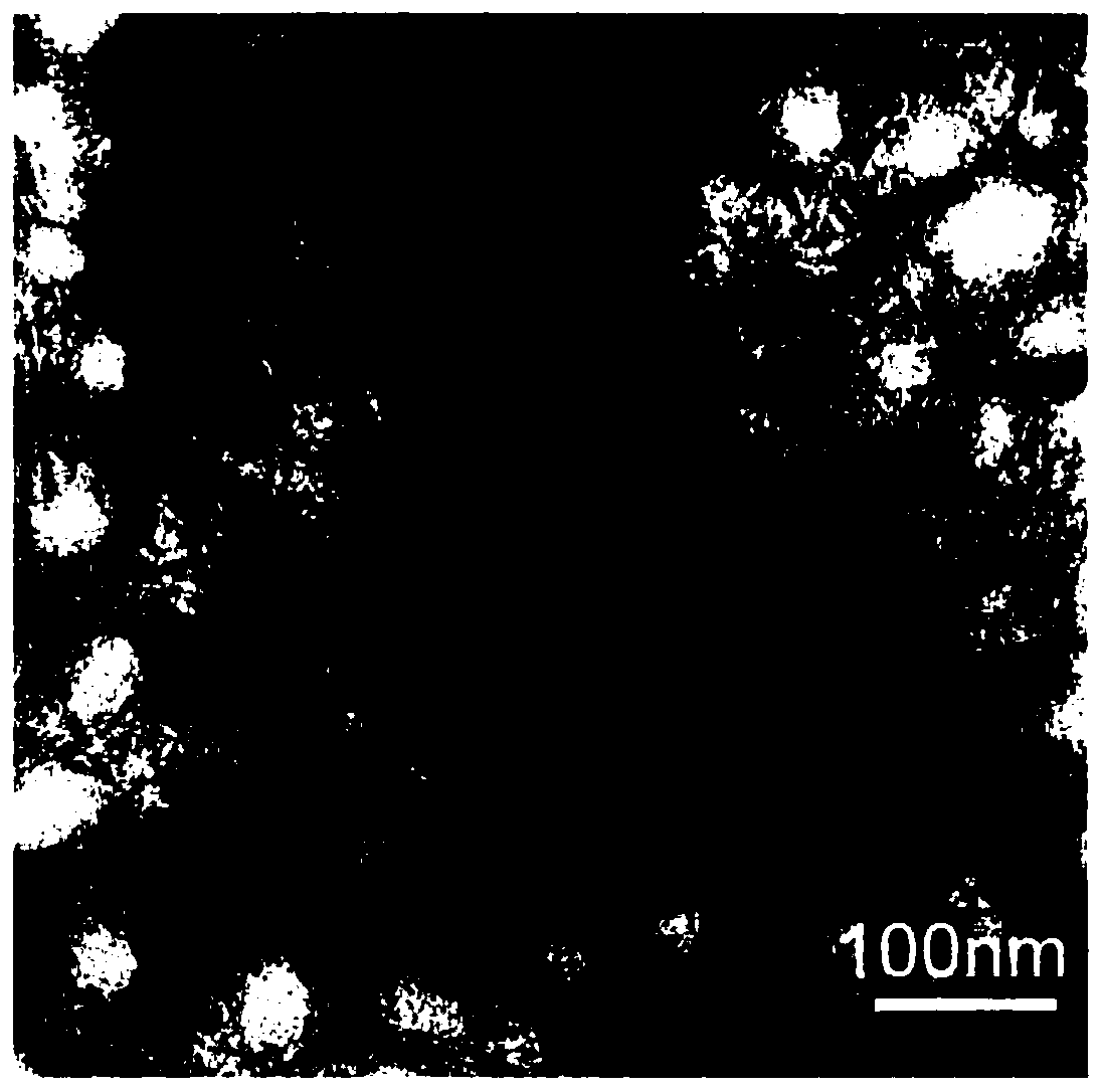
[0043] HPV16 E7 44-62 The peptide was chemically modified to the N-terminus of the self-assembling peptide Q11 through the flexible arm Ser-Gly-Ser-Gly, and the E7-Q11 peptide was purified by reverse-phase HPLC, lyophilized, and stored at -20°C. Dissolve lyophilized E7-Q11 in sterile water to 2mM, and place it at 4°C overnight, then assemble E7-Q11 into nanofibers with 1×PBS (pH7.4) solution, the assembly condition is 1×PBS (pH7.4 ) solution to dilute the E7-Q11 aqueous solution to 0.5 mM, and stand at room temperature 20° C. for 4.5 hours to obtain an anti-HPV-related tumor vaccine assembled into nanofibers.
[0044] The assembled E7-Q11 nanofibers were observed by a transmission electron microscope (TEM) for the morphology of the assembled E7-Q11 fibers.
[0045] Among them, Q11 peptide (amino acid sequence: Ac-Gln-Gln-Lys-Phe-Gln-Phe-Gln-Phe-Glu-Gln-Gln-Am, such as SED IQ NO.4...
Embodiment 2
[0051] Example 2: Preventive immunization strategy and mouse transplantation tumor model establishment
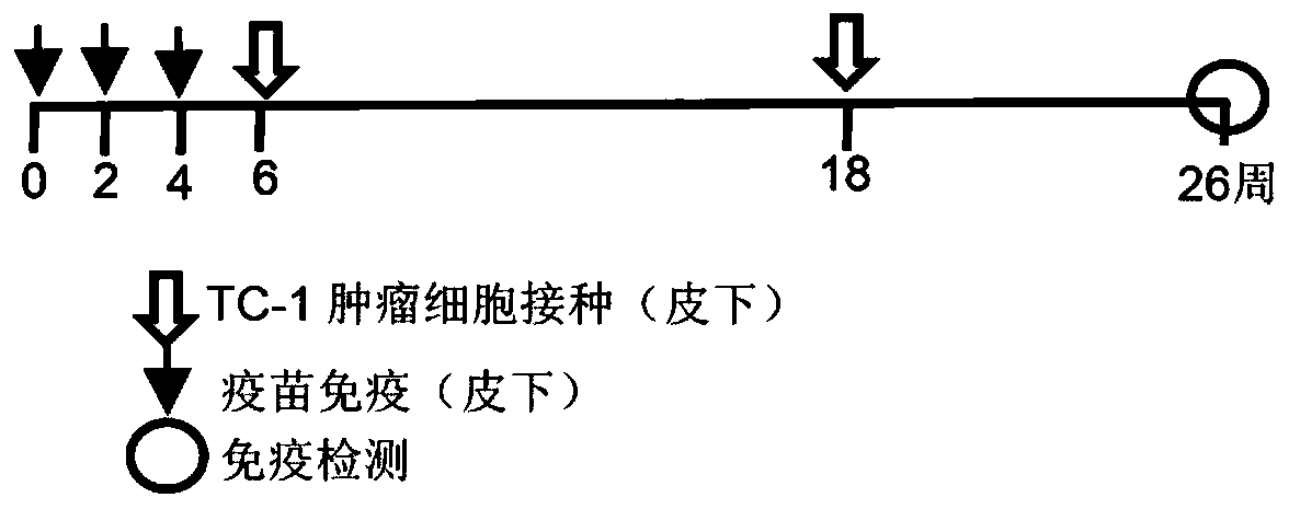
[0052] Female C57BL mice, 6-8 weeks old, in the preventive immunization experiment, the mice were first immunized 3 times with an interval of 2 weeks, and the mice were divided into two groups, with 5 mice in each group, which were the immune Q11 carrier group ( Using Q11 peptide) and E7-Q11 vaccine group (using nanofiber E7-Q11), the immunological dose is 12.5nmol, each 200ul, two weeks after the third immunization, subcutaneously inject a volume of 50ul in the right abdomen of the mouse, 1 ×10 5 TC-1 cells (cell concentration 2×10 6 cells / ml), and the volume ratio of cells and Basement MembraneMatrix is 1:1 ( image 3 ). After tumor inoculation, tumor size was measured every 3-4 days ( Figure 4 ). Twelve weeks after the first tumor inoculation, the tumor was re-inoculated with 1 × 10 subcutaneously in the left abdomen. 5 TC-1 cells (cell concentration 2×10 6 cel...
Embodiment 3
[0053] Example 3: Establishment of transplanted tumor models in mice and therapeutic immunity to 2-3mm tumors
[0054] The mice were divided into 3 groups, and 1 × 10 5 TC-1 cells (cell concentration 2×10 6 cells / ml), immunization procedures such as Figure 5 , when the tumor grows to 2-3mm, the vaccine is immunized, and the mice are divided into three groups, namely the Q11 vector group, the E7-Q11 vaccine group, and the E7-Q11 non-assembly group (the E7-Q11 non-assembly group is E7-Q11 Immediately immunize mice with lyophilized peptide dissolved in sterile water (without 1×PBS assembly), the immunological dose is 12.5nmol, 100ul each time, immunized 3 times in total, with one week interval, and the size of the tumor is measured every 3-4 days ( Figure 6 , Figure 7 ), 34 days after the third immunization, the spleen of the mice was taken for immunological detection, using ELISPOT to detect antigen-specific lymphocytes secreting IFN-γ, and using E7 49-57 Peptides were u...
PUM
| Property | Measurement | Unit |
|---|---|---|
| control rate | aaaaa | aaaaa |
Abstract
Description
Claims
Application Information
 - R&D
- Intellectual Property
- Life Sciences
- Materials
- Tech Scout
- Unparalleled Data Quality
- Higher Quality Content
- 60% Fewer Hallucinations
Browse by: Latest US Patents, China's latest patents, Technical Efficacy Thesaurus, Application Domain, Technology Topic, Popular Technical Reports.
© 2025 PatSnap. All rights reserved.Legal|Privacy policy|Modern Slavery Act Transparency Statement|Sitemap|About US| Contact US: help@patsnap.com


