Eureka AIR delivers breakthrough ideas for toughest innovation challenges, trusted by R&D personnel around the world.

Method for inducing human umbilical cord derived mesenchymal stem cells to differentiate into chondrocytes and application thereof

A technology of mesenchymal stem cells and chondrocytes, applied in the field of biomedicine, can solve the problems that affect the clinical application of adipose MSCs, the quality of cells is affected by the donor, and the limited proliferation ability, so as to improve the potential of multidirectional differentiation, promote the ability of mineralization and differentiation, The effect of promoting stem cell differentiation

Pending Publication Date: 2019-09-06
WUHAN HAMILTON BIOTECH
View PDF2 Cites 0 Cited by
  • Summary
  • Abstract
  • Description
  • Claims
  • Application Information

AI Technical Summary

Problems solved by technology

However, autologous bone joint or bone marrow stem cells from patients with severe osteoarthritis often have very limited proliferative ability
Regardless of autologous or allogeneic bone marrow MSCs, there are defects such as traumatic extraction, low output, and cell quality affected by the donor.
The same problem also affects the clinical application of adipose MSCs

Method used


Image

  • Method for inducing human umbilical cord derived mesenchymal stem cells to differentiate into chondrocytes and application thereof
  • Method for inducing human umbilical cord derived mesenchymal stem cells to differentiate into chondrocytes and application thereof
  • Method for inducing human umbilical cord derived mesenchymal stem cells to differentiate into chondrocytes and application thereof

Examples


Embodiment 1

[0052] S1. Resuscitating UC-MSCs cells: take the UC-MSCs to be tested, quickly recover at 37°C, and resuspend in complete medium;

[0053] Preparation of cell microspheres: collect recovered UC-MSCs and dilute 1×10 7 Cells / mL were resuspended in complete medium, and the cell suspension was added dropwise to the bottom of a 6-well cell culture plate (15L / drop, 5-7 drops / well), and transferred to a cell culture incubator with 5% CO 2 , Culture at 37°C for 2 hours, then add 2 mL of complete medium to each well, continue culturing overnight until the cell droplets shrink to form cell microspheres, and observe under a microscope during the culture process (such as Figure 11 , 12 shown);

[0054] S2. The medium for culturing the cell microspheres is replaced with a chondrogenic differentiation medium, the medium is replaced every 3 days, and the culture is sufficient for 14-28 days.

Embodiment 2

[0055] Embodiment two is comparative example

[0056] The difference from Example 1 is:

[0057] Observing the structure under a microscope during the cultivation process in step S1 is as follows: Figures 13 to 17 shown;

[0058] In S2, after the cell microspheres are formed, the complete medium is continued to be used for culturing, and the medium is replaced every other day for 14-28 days.

[0059] The following setup test detects related product properties etc. in the method of the present invention;

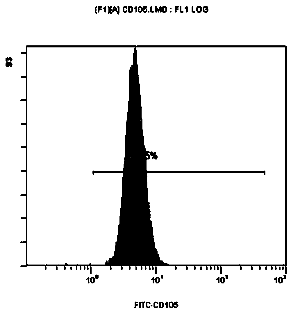
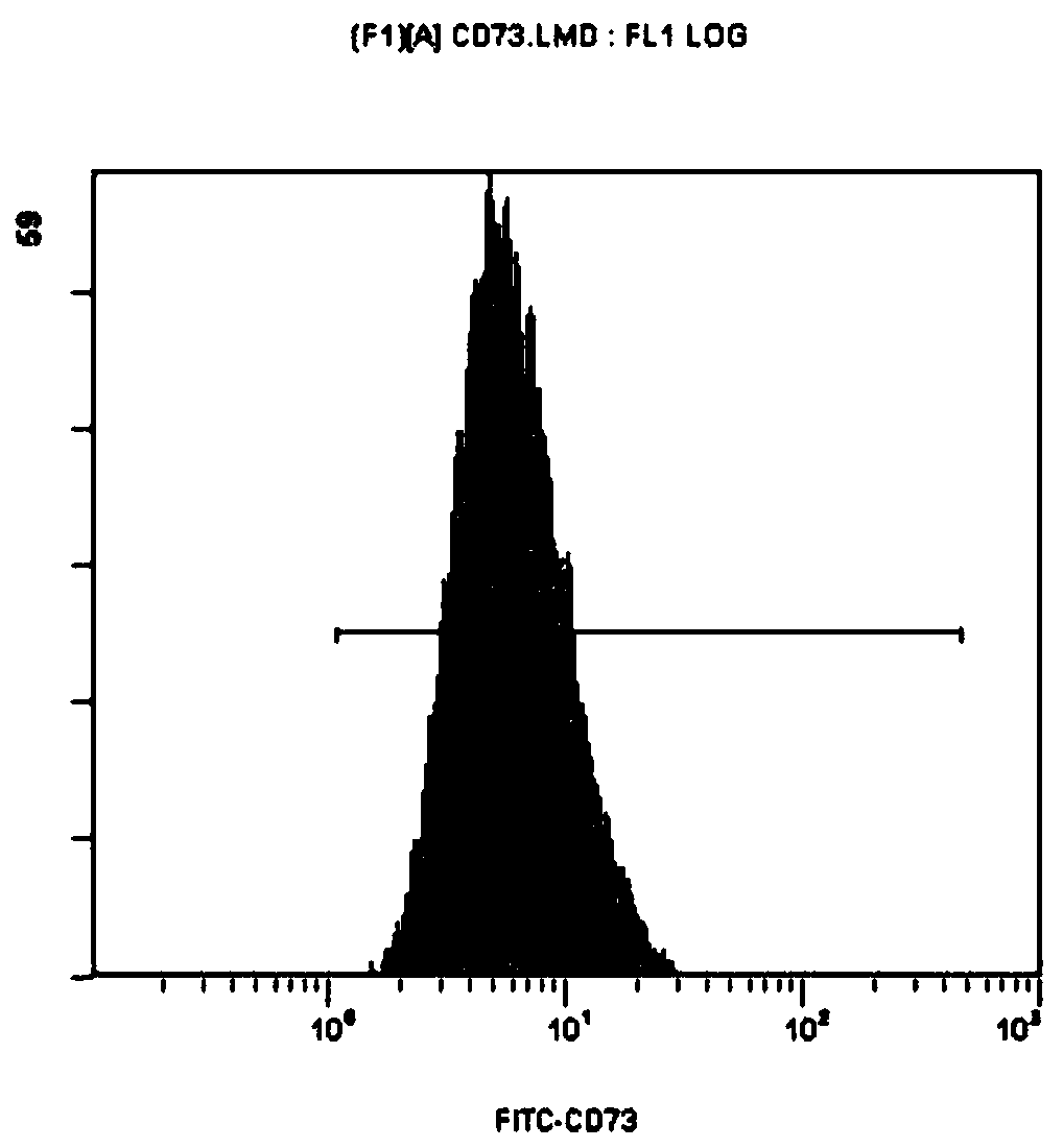
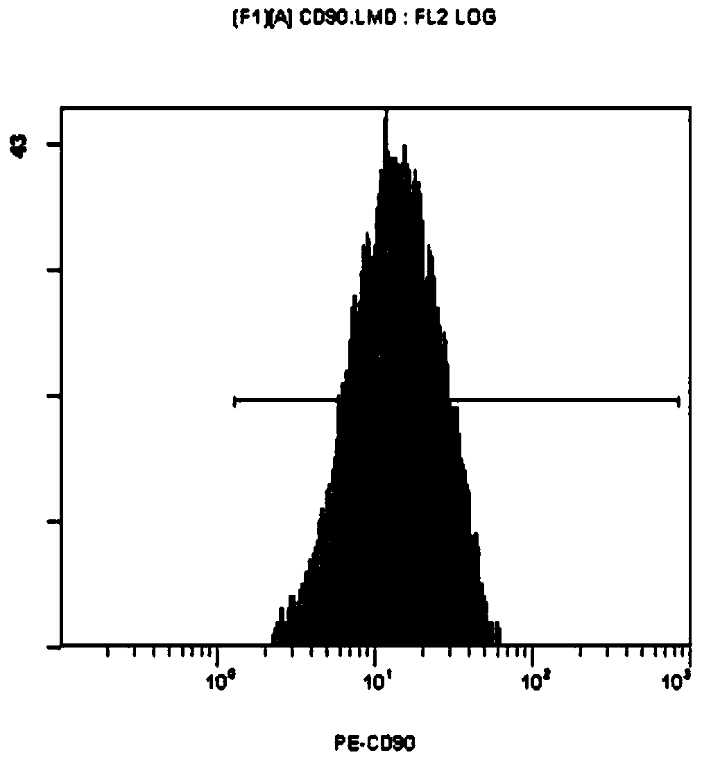
[0060] 1. Identification of UC-MSCs immunolabeling:

[0061] Culture the UC-MSCs obtained in step S1 to collect the 5th passage UC-MSCs. After washing with PBS, prepare 6 × 10 6 cells / mL of cell suspension; press the cell suspension to 100μL / tube (5×10 5 Cells) were aliquoted, and 5 μL of CD105, CD90, CD44, CD73, CD19, CD34, CD45 and HLA-DR monoclonal antibodies labeled with different fluoresceins were added, mixed slightly by shaking, and incubated at room temperature ...


PUM


Abstract

A method for inducing human umbilical cord derived mesenchymal stem cells to differentiate into chondrocytes is characterized by including the steps: S1, resuscitating UC-MSCs, collecting the resuscitated UC-MSCs and preparing cell microspheres by a 3D pendant-drop method; and S2, replacing a culture medium for culturing the cell microspheres with a cartilage-induced differentiation culture medium, replacing the culture medium once every other 3 days, and culturing for 14-28 days. The method has the advantages: 1, in the preparation method, the UC-MSCs are prepared in a three-dimensional three-dimensional environment, so the contact between cells is broader and cell bridging and close connection and formation of cholangiole are facilitated, so as to maintain the UC-MSCs phenotype stability; and 2, 3D pendant-drop culture is adopted, so that cell arrangement is closer, changes in cell micro-environments makes a series of signal transduction and intracellular signal transduction betweenstem cell surface membrane molecules changed, so as to promote the differentiation of various stem cells.

Description

technical field [0001] The invention relates to the field of biomedicine, regenerative medicine and bone disease technology, in particular to a method and application field for inducing human umbilical cord-derived mesenchymal stem cells to differentiate into chondrocytes. Background technique [0002] Osteoarthritis (OA) is a chronic inflammatory disease of bones and joints, characterized by degeneration and destruction of articular cartilage, subchondral bone sclerosis and osteophyte formation, and inflammation and hyperplasia of surrounding soft tissues. joints, such as knee osteoarthritis (Knee osteoarthritis, KOA). There are about 1 billion OA patients in the world, of which about 100 million are Chinese patients. The incidence of OA increases with age. Preliminary estimates suggest that the prevalence of OA in my country is about 62% in people over 60 years old, and as high as 80% in people over 75 years old. The clinical manifestation is repeated and persistent bone...

Claims


Application Information

Patent Timeline
Patent Type & Authority Applications(China)
IPC IPC(8): C12N5/077C12N5/0775
CPCC12N5/0655C12N2506/1369C12N2513/00C12N2500/90C12N2500/32C12N2500/38C12N2527/00C12N2500/30
Inventor 张权武栋成
Owner WUHAN HAMILTON BIOTECH
Why Eureka
Eureka Blog
Learn More