Magnetic nanoparticle bone repair scaffold and preparation method thereof

A magnetic nanometer and magnetic nanoparticle technology, applied in the field of biomedical engineering, can solve the problems of postoperative complications of secondary injury, limited source of autologous bone, immune rejection, etc. Effects of blood vessel formation and bone formation promotion

Inactive Publication Date: 2019-09-13
PEKING UNION MEDICAL COLLEGE HOSPITAL CHINESE ACAD OF MEDICAL SCI +1
View PDF7 Cites 3 Cited by
  • Summary
  • Abstract
  • Description
  • Claims
  • Application Information

AI Technical Summary

Problems solved by technology

Although the therapeutic effect of autologous bone grafting is good, the source of autologous bone is limited, and there is a risk of secondary injury and postoperative complications
However, if allogeneic bone transplantation is used, although the source of allogeneic bone is sufficient, it will cause immune rejection and there is a risk of potential disease infection

Method used


Image

  • Magnetic nanoparticle bone repair scaffold and preparation method thereof
  • Magnetic nanoparticle bone repair scaffold and preparation method thereof
  • Magnetic nanoparticle bone repair scaffold and preparation method thereof

Examples


Embodiment 1

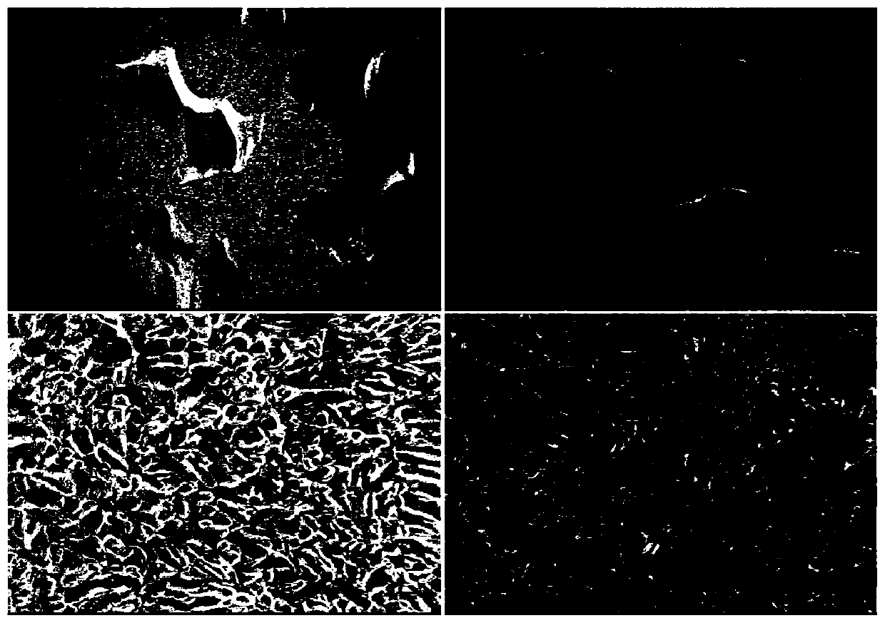
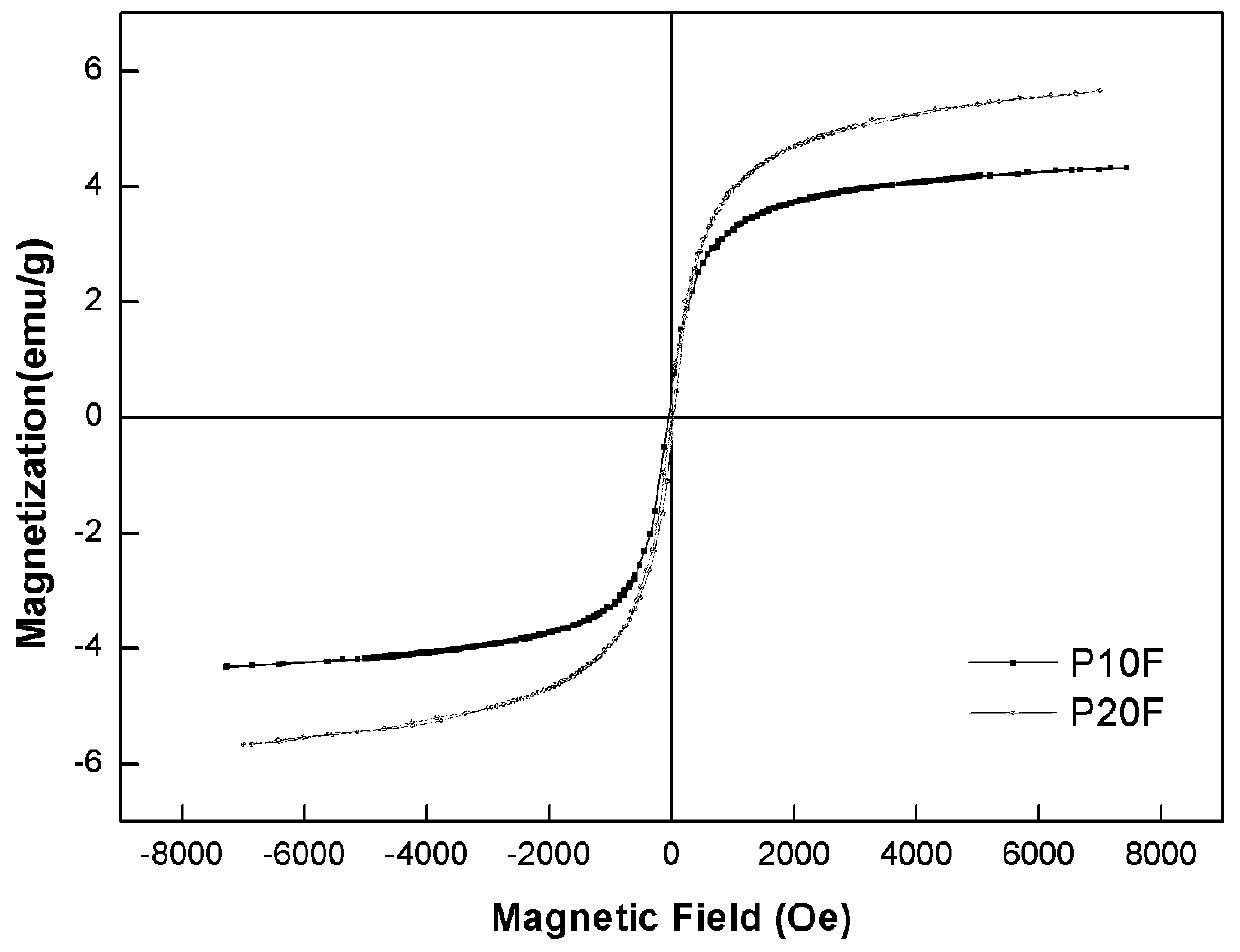
[0030] The magnetic nano-bone repair scaffold material of the present embodiment is made up of the following mass percentage components: 90% PLGA and 10% γ-Fe 2 o 3 nanoparticles.

[0031]The preparation method of the above-mentioned magnetic nano-bone repair scaffold comprises the following steps:

[0032] 1) According to the mass percentage, weigh 90% of PLGA and 10% of the nanoparticles with a particle size of 10nm and place them in a flask, then add 1,4-dioxane, and stir overnight at room temperature To form a homogeneous solution, wherein the concentration of PLGA in 1,4-dioxane is 0.14g / mL;

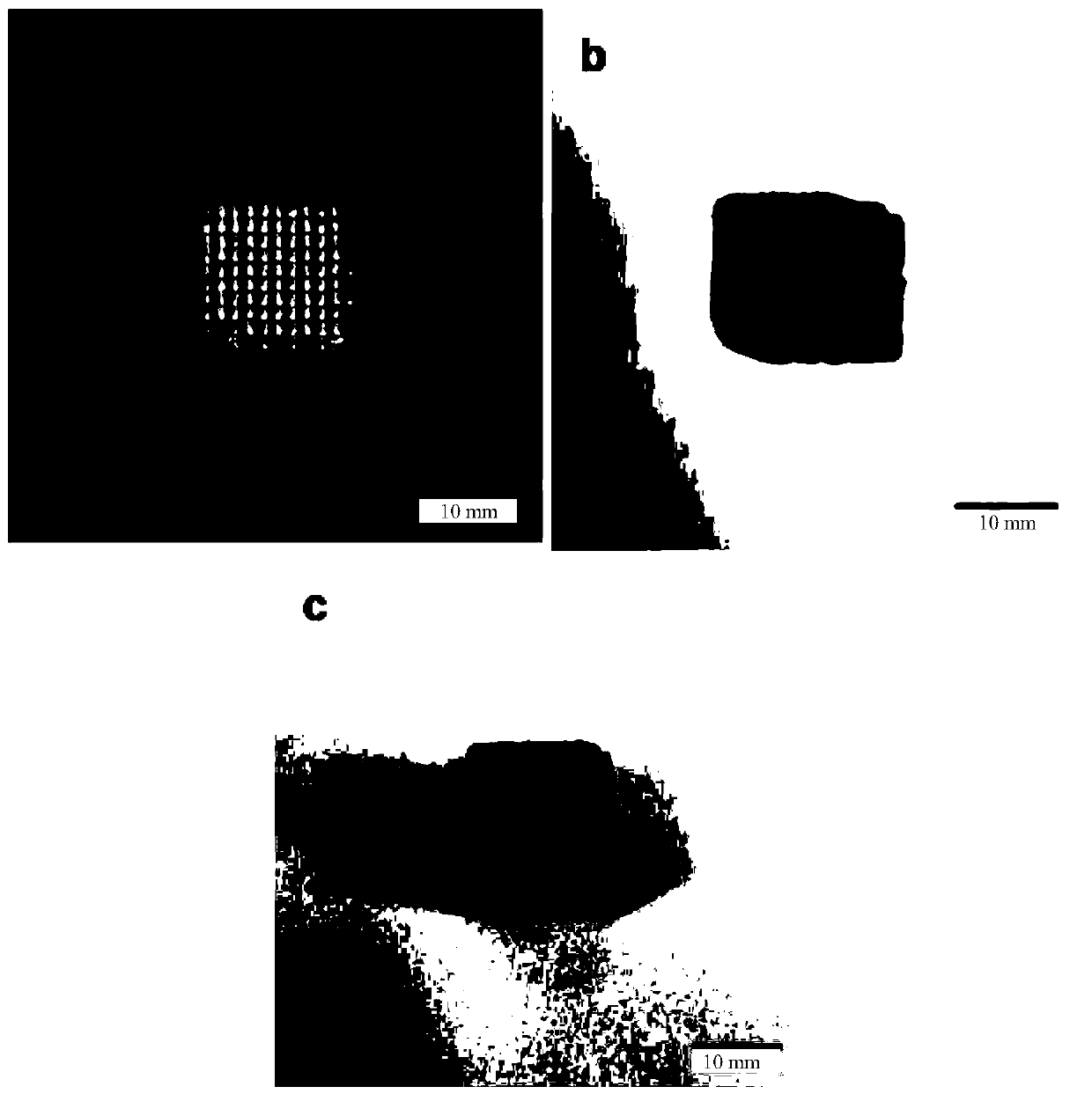
[0033] 2) Use SolidWorks software to design and create a model, for example, 1.4×1.4×1.4cm 3 The cube structure model, and export the data containing the SolidWorks model, and then use the layering software to process the data containing the SolidWorks model layered to obtain the layered data, and generate a CLI file;

[0034] 3) Add the homogeneous solution obtained in step 1) ...

Embodiment 2

[0037] The magnetic nano-bone repair scaffold material of the present embodiment is made up of the following mass percentage components: 80% PLGA and 20% γ-Fe 2 o 3 nanoparticles.

[0038] The preparation method of the above-mentioned magnetic nano-bone repair scaffold comprises the following steps:

[0039] 1) According to the mass percentage, weigh 80% of PLGA and 20% of the nanoparticles with a particle size of 10nm and place them in a flask, then add 1,4-dioxane, and stir overnight at room temperature To form a homogeneous solution, wherein the concentration of PLGA in 1,4-dioxane is 0.14g / mL;

[0040] 2) Use SolidWorks to design and create a model, for example, 1.4×1.4×1.4cm 3 The cube structure model, and export the data containing the SolidWorks model, and then use the layering software to process the data containing the SolidWorks model layered to obtain the layered data, and generate a CLI file;

[0041] 3) Add the homogeneous solution obtained in step 1) into the...

Embodiment 3

[0044] The method of this embodiment is basically the same as that of Example 1, except that the prepared magnetic nano-bone repair scaffold is composed of the following components by mass percentage: 95% PLGA, 5% γ-Fe 2 o 3 nanoparticles.


PUM

PropertyMeasurementUnit
Apertureaaaaaaaaaa
Particle sizeaaaaaaaaaa
Apertureaaaaaaaaaa

Abstract

The invention discloses a biodegradable magnetic nanoparticle bone repair scaffold and a preparation method thereof. The biodegradable magnetic nanoparticle bone repair scaffold of the invention is aporous scaffold prepared by three-dimensional printing after software modeling, so that bone repair scaffolds of different structures can be designed according to bone defect shapes; the biodegradablemagnetic nanoparticle bone repair scaffold contains nanoparticles with superparamagnetism under the magetostatic field, so that the biodegradable magnetic nanoparticle bone repair scaffold is good inbiocompatibility and capable of being degraded in vivo and playing a role in promoting osteogenesis; the nanoparticles produce Fe 3+(superscript) in the degradation process, and the Fe 3+(superscript) can promote expressions of angiogenic factors in HUVEC, so that the biodegradable magnetic nanoparticle bone repair scaffold has an excellent function of promoting angiogenesis; and the scaffold hasrelatively high compression modulus, good superparamagnetism, and high porosity, so that the scaffold is conducive to the growth, adhesion and proliferation of osteoblasts. Therefore, as a bone repair scaffold to guide bone regeneration, the scaffold has great potential application value in the field of bone defect treatment.

Description

technical field [0001] The invention relates to the technical field of biomedical engineering, in particular to a magnetic nano-bone repair bracket and a preparation method thereof. Background technique [0002] Nowadays, bone defects caused by aging, trauma, and bone disease are difficult problems in the current orthopedic surgery. At present, autologous bone grafting is mostly used in the clinical field to repair local bone defects. Although the therapeutic effect of autologous bone grafting is good, the sources of autologous bone are limited, and there are risks of secondary injury and postoperative complications. However, if allogeneic bone transplantation is used, although the source of allogeneic bone is sufficient, it will cause immune rejection and there is a risk of potential disease infection. Therefore, the use of multiple biocompatible materials is currently a research hotspot in tissue engineering scaffolds. Through the combination of different materials, a b...

Claims


Application Information

Patent Timeline
IPC IPC(8): A61L27/18A61L27/02A61L27/50A61L27/54A61L27/56A61L27/58B33Y80/00
CPCA61L27/025A61L27/18A61L27/50A61L27/54A61L27/56A61L27/58A61L2300/412A61L2400/12A61L2430/02B33Y80/00C08L67/04
Inventor 王海刘洁颖赖毓霄郑子卓吴志宏邱贵兴
Owner PEKING UNION MEDICAL COLLEGE HOSPITAL CHINESE ACAD OF MEDICAL SCI
Why Eureka
Try Eureka