Living body tracing cerebral nerve connection method based on magnetic resonance imaging and application thereof

A magnetic resonance imaging and brain technology, applied in the biological field, can solve problems such as the inability to display neuron connection information, and achieve the effects of flexible change, high sensitivity, and clear connection progression.

Pending Publication Date: 2019-11-15
WUHAN INST OF PHYSICS & MATHEMATICS CHINESE ACADEMY OF SCI
View PDF3 Cites 4 Cited by
  • Summary
  • Abstract
  • Description
  • Claims
  • Application Information

AI Technical Summary

Problems solved by technology

However, since adenovirus can only infect local neurons at the injection site, it cannot spread across synapses between neurons and nerves, so the connection information between neurons cannot be displayed

Method used


Image

  • Living body tracing cerebral nerve connection method based on magnetic resonance imaging and application thereof
  • Living body tracing cerebral nerve connection method based on magnetic resonance imaging and application thereof
  • Living body tracing cerebral nerve connection method based on magnetic resonance imaging and application thereof

Examples


Embodiment 1

[0025] Example 1: Construction scheme of recombinant AAV of rAAV2-retro serotype carrying Ferritin gene

[0026] Schematic diagram of rAAV2-retro-CAG-Ferritin-WPRE-pA genome ( figure 1 ), insert CAG promoter (shown in SEQ ID NO. 1), Ferritin gene (shown in SEQ ID NO. 2), WPRE post-transcriptional regulatory element (shown in SEQ ID NO. 3) and polyA sequence (shown in SEQ ID NO. Shown in NO.4). This design scheme was handed over to Wuhan Secret Brain Science and Technology Co., Ltd. for the subsequent construction of rAAV2-retro serotype recombinant AAV virus to obtain recombinant virus rAAV2-retro-CAG-Ferritin-WPRE-pA.

Embodiment 2

[0027] Example 2: Immunohistochemical detection and MRI observation of rAAV2-retro-CAG-Ferritin-WPRE-pA injected into the brain of mice

[0028] The rAAV2-retro-CAG-Ferritin-WPRE-pA virus (constructed by Wuhan Central Brain Science and Technology Co., Ltd.) was injected into the dorsal striatal brain area of ​​mice (CPu, coordinates: AP-0.5mm; ML-2mm; DV-3.3mm), its titer is 5.57×10 12 vg / ml, the injection volume is 2.8μL.

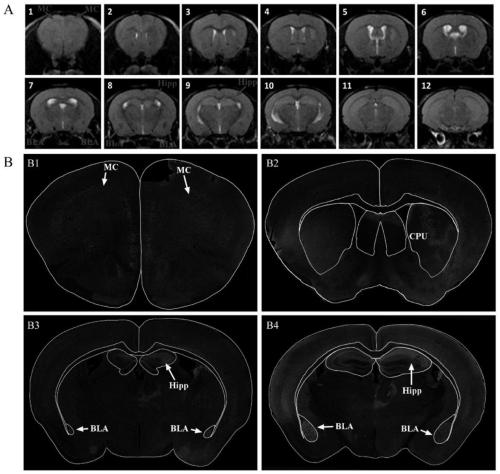
[0029] After 40 days, the animal brain was observed in vivo using a 7.0T magnetic resonance imaging instrument. The T2-weighted imaging sequence used was the RARE relaxation enhancement rapid acquisition sequence. The parameters were: TR=3000ms, effTE=55ms, Rarefactor=4 , The total duration of the sequence is 28min. The FOV is 1.75×1.75cm, the resolution is 0.136×0.136mm, and the layer thickness is 0.5mm. MRI results ( figure 2 A) It shows that the virus successfully enters the neuron, and enters the neuron cell body (lateral amygdala BLA, hippocampus Hipp, ...

Embodiment 3

[0032] Example 3: Long-term MRI observation of rat brain in vivo at different time points after rAAV2-retro-CAG-Ferritin-WPRE-pA injection

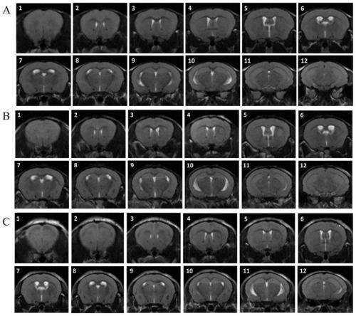
[0033] One of the advantages of the neural connection tracking method proposed by the present invention is that the brain neural connection can be observed in vivo for a long time. Therefore, in this example, the brain MRI results of the same mouse before rAAV2-retro-CAG-Ferritin-WPRE-pA virus injection, 30 days after injection and 70 days after injection are given. Before virus injection, the animal brain was observed in vivo with a 7.0T magnetic resonance imaging instrument. The T2-weighted imaging sequence used was the RARE relaxation enhancement rapid acquisition sequence. The parameters were: TR=3000ms, effTE=55ms, Rarefactor =4, the total duration of the sequence is 28min. The FOV is 1.75×1.75cm, the resolution is 0.136×0.136mm, and the layer thickness is 0.5mm. Then the rAAV2-retro-CAG-Ferritin-WPRE-pA virus was injected into the do...


PUM


Abstract

The invention discloses a living body tracing cerebral nerve connection method based on magnetic resonance imaging and application thereof. The method comprises the steps of using an adeno-associatedvirus variant rAAV2-retro with an axon terminal absorption reverse transport characteristic as a carrier, loading a gene of Ferritin in a genome of the carrier, packaging the carrier into a recombinant virus rAAV2-retro-CAG-Ferritin-WPRE-pA, injecting the recombinant virus into the brain, infecting neurons with the virus, expressing Ferritin in the neurons, and observing nerve connection between other brain regions and the infected neurons at an injected site in combination with MRI. The method combines magnetic resonance imaging with rAAV2-retro-CAG-Ferritin-WPRE-pA, realizes living body tracing of cerebral nerve connection and can be used for long-time nerve structure loop tracing or nerve function analysis.

Description

Technical field [0001] The invention belongs to the field of biotechnology, and specifically relates to a method for tracing brain nerve connections in a living body based on magnetic resonance imaging, and also relates to the application of the method in neural structure loop tracing and functional analysis. Background technique [0002] The neural network of the brain performs important functions in the body, participating in various activities such as learning, memory, cognition and emotion, and a variety of neurological diseases are closely related to the abnormality of the neural network. Analyzing the neural structure network of the brain plays an important role in understanding the brain's function, working mechanism, development, degeneration, and the occurrence and development of neurological diseases. In recent years, neurotropic viruses, such as rabies virus, vesicular stomatitis virus, herpes simplex virus, pseudorabies virus, etc., have been widely used in neural net...

Claims


Application Information

Patent Timeline
Patent Type & Authority Applications(China)
IPC IPC(8): C12N15/861A61K49/18
CPCC12N15/86A61K49/1896C12N2710/10043
Inventor 郑宁张志建何晓斌苏鹏林坤章蔡奥灵卢晨王杰徐富强
Owner WUHAN INST OF PHYSICS & MATHEMATICS CHINESE ACADEMY OF SCI
Why Eureka
Try Eureka