Biosensor for detecting PS exosome and preparation method of biosensor

A biosensor and biosensing technology, applied in the direction of chemiluminescence/bioluminescence, instruments, measuring devices, etc., can solve the problems of lack of stability, denaturation, low sensitivity, etc., and achieve the effect of improving selectivity and stability

Inactive Publication Date: 2019-11-15
BEIJING UNIV OF CHEM TECH
View PDF0 Cites 2 Cited by
  • Summary
  • Abstract
  • Description
  • Claims
  • Application Information

AI Technical Summary

Problems solved by technology

[0006] The purpose of the present invention is to provide a biosensor for detecting PS-positive tumor-derived exosomes and a preparation method, by identifying the highly specific binding of peptide-based probes to PS-positive exosomes, realizing the detection of PS-positive exosomes in samples Perform rapid and highly sensitive detection to solve

Method used


Image

  • Biosensor for detecting PS exosome and preparation method of biosensor
  • Biosensor for detecting PS exosome and preparation method of biosensor
  • Biosensor for detecting PS exosome and preparation method of biosensor

Examples


Embodiment 1

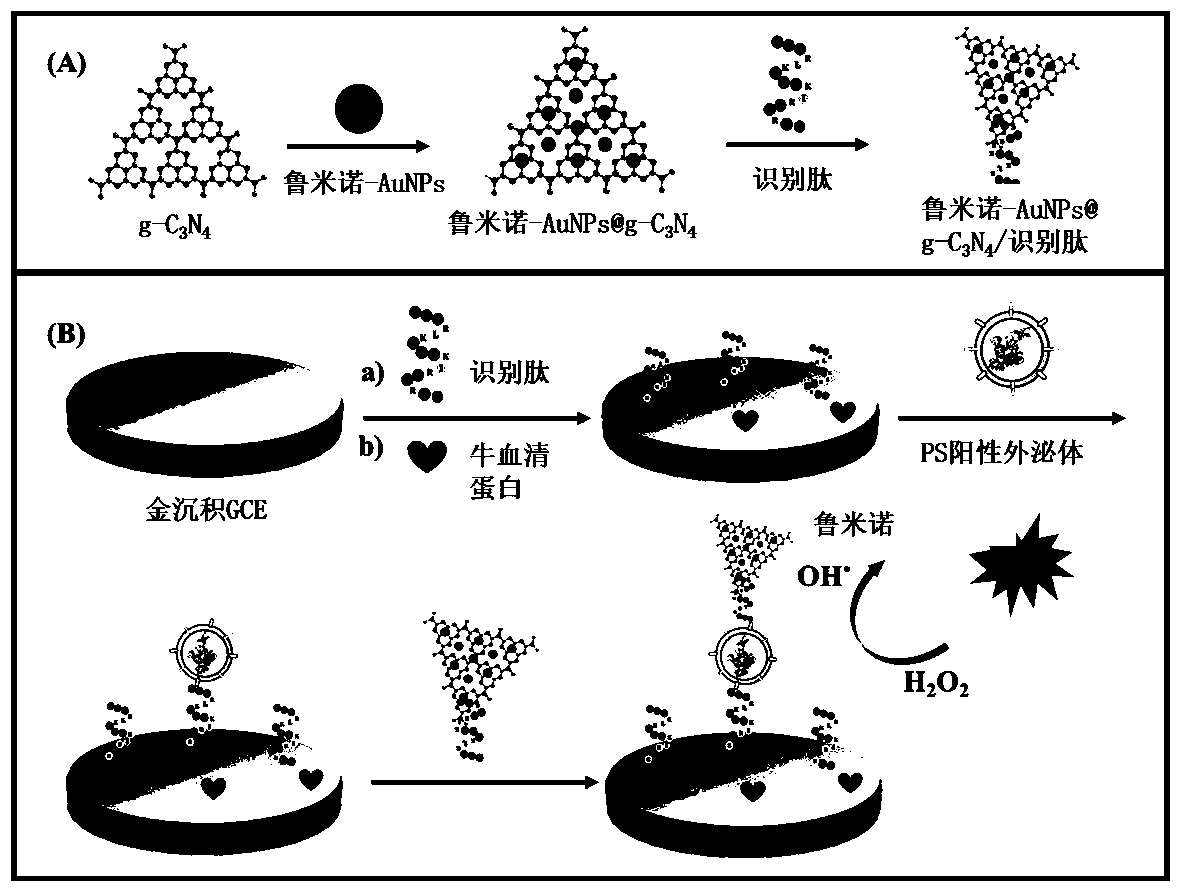
[0026] 1. g-C 3 N 4 Synthesis of Nanosheets and Luminol-AuNPs

[0027] Preparation of g-C by direct pyrolysis of melamine 3 N 4 . The melamine was heated at 550°C for 4 hours, held at this temperature for a further 4 hours and cooled to room temperature. The resulting yellow product is then lumpy g-C 3 N 4 powder. Synthesize g-C as follows 3 N 4 Nanosheets: to 1 g of the bulk g-C 3 N 4 Add 100 mL 5mol / L HNO to the powder 3 , reflux at 125 °C for 24 h, and cool at room temperature. The white product was centrifuged at 12000 rpm, washed with ultrapure water to near neutral pH, and redispersed in water. To obtain uniformly sized nanosheets, the resulting suspension was sonicated for 4 h and then centrifuged at 8000 rpm for 30 min to remove residual unexfoliated g-C 3 N 4 Nanoparticles and large-area nanosheets. Then, the obtained supernatant was centrifuged at 5000 rpm to remove the suspension. Finally, the product was dried in a vacuum oven at 35 °C for 12 hours...

Embodiment 2

[0037] 1. Luminol-AuNPs@g-C 3 N 4 Synthesis of / PS recognition peptide probes

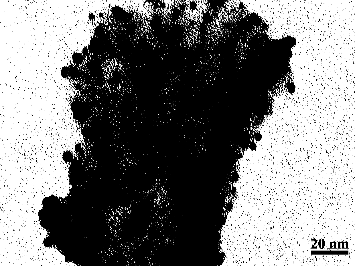
[0038] 100 mg g-C 3 N 4 After mixing the nanosheets and 50 mL luminol-AuNPs solution evenly, stir at room temperature for 18 h, and then centrifuge at 6000 rpm for 30 minutes to remove the supernatant to obtain luminol-AuNPs@g-C 3 N 4 Precipitate, figure 2 For Luminol-AuNPs@g-C 3 N 4 Transmission electron microscope image of a nanomaterial. The resulting luminol-AuNPs@g-C 3 N 4 Luminol-AuNPs@g-C can be obtained by combining the precipitate with the recognition peptide through thiol-gold coordination at 30°C for 6 h 3 N 4 / PS recognizes a peptide signaling probe.

[0039] 2. Assembly of the biosensor

[0040] Using 1% chloroauric acid solution as the reaction electrolyte, AuNPs were modified onto the glassy carbon electrode by potentiostatic deposition at -0.2 V for 30 s, rinsed and dried; then the modified electrode was immersed in 0.5 μM recognition peptide solution4 Incubate at ℃ f...

Embodiment 3

[0044] 1. Luminol-AuNPs@g-C 3 N 4 Synthesis of / PS recognition peptide probes

[0045] 50 mg g-C 3 N 4 After mixing the nanosheets and 100 mL luminol-AuNPs solution evenly, stir at room temperature for 36 h, and then centrifuge at 6000 rpm for 30 min to remove the supernatant to obtain luminol-AuNPs@g-C 3 N 4 Precipitate, figure 2 For Luminol-AuNPs@g-C 3 N 4 Transmission electron microscope image of a nanomaterial. The resulting luminol-AuNPs@g-C 3 N 4 Luminol-AuNPs@g-C can be obtained by combining the precipitate and the recognition peptide through the coordination of thiol and gold at 40°C for 8 h 3 N 4 / PS recognizes a peptide signaling probe.

[0046] 2. Assembly of the biosensor

[0047] Using 1% chloroauric acid solution as the electrodeposition base solution, AuNPs were modified on the glassy carbon electrode by constant potential deposition at -0.2 V for 20 s, rinsed and dried; then the modified electrode was immersed in 1 μM recognition peptide solution4...


PUM


Abstract

The invention provides a biosensor for a phosphatidyl serine (PS) exosome and a preparation method of the biosensor. According to the method, a PS specific recognition peptide serves as a recognitionmodule, a biosensing electrode assembled by the recognition peptide serves as a capture substrate, and an electrochemiluminescence probe formed by a g-C3N4 nanosheet, luminol-AuNPs and the recognitionpeptide serves as a signal probe. The PS exosome in the sample is specifically recognized by the capture substrate and the signal probe in order to form a sandwich structure, and a detection signal is generated through the signal probe. The PS recognition peptide can be specifically combined with the PS on the exosome of a tumor source, and the PS recognition peptide is simple in structure, highin affinity and selectivity, not prone to be influenced by environmental factors such as pH and temperature, and good in stability. According to the biosensor and the preparation method of the biosensor, high-sensitivity and high-selectivity PS exosome detection can be realized.

Description

technical field [0001] The invention belongs to the technical field of exosome detection, and in particular relates to an electrochemiluminescent sensor for detecting tumor-derived exosomes based on a recognition peptide and a preparation method thereof. Background technique [0002] Cancer has become the number one killer threatening human health, and its early detection and accurate detection have very important practical significance for reducing patient mortality and improving survival rate. For a long time, tumor tissue biopsy has often been used as the gold standard for tumor diagnosis. However, with the deepening of tumor research, scientists have found that tissue biopsy techniques have certain limitations in the process of cancer diagnosis and treatment. The main manifestation is that the tumor is heterogeneous, some patients are not suitable for tissue biopsy, and the hysteresis of tissue biopsy is not good for the treatment of patients. Tumor marker detection has...

Claims


Application Information

Patent Timeline
IPC IPC(8): G01N33/574G01N21/76G01N27/327
CPCG01N21/76G01N27/327G01N33/57449
Inventor 陈旭刘雪娇杨文胜
Owner BEIJING UNIV OF CHEM TECH
Why Eureka
Try Eureka