
Reinforcing method for early embryo optical coherence tomography image contrast

An optical coherence tomography and image comparison technology, applied in the field of biomedical imaging, can solve the problems of limited applicability of mammalian embryo blood vessel remodeling, expensive imaging system, etc., to improve experimental data reference and research strategies, easy operation, The test results are obvious

Active Publication Date: 2019-11-22
XIAMEN UNIV
View PDF0 Cites 0 Cited by
  • Summary
  • Abstract
  • Description
  • Claims
  • Application Information

AI Technical Summary

Problems solved by technology

Larin et al. used swept-frequency optical coherence tomography to study the normal vascular development of zebrafish and chicken embryos, but the applicability to mammalian embryo vascular remodeling is limited, and the imaging system is expensive

Method used


Image

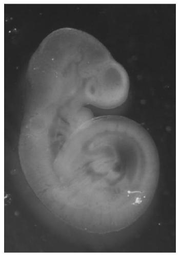
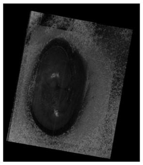
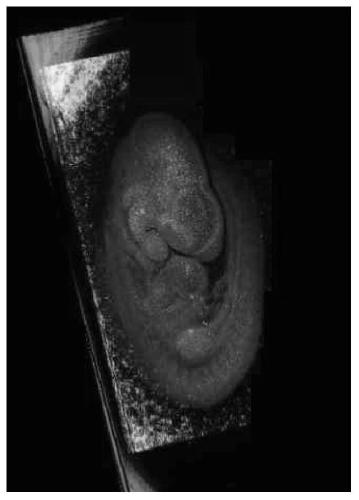
  • Reinforcing method for early embryo optical coherence tomography image contrast
  • Reinforcing method for early embryo optical coherence tomography image contrast
  • Reinforcing method for early embryo optical coherence tomography image contrast

Examples


Embodiment 1

[0029] A method for enhancing the contrast of an early embryo optical coherence tomography image, comprising the following steps:

[0030] (1) Separate early embryos with complete placenta and vitelline membrane from the mother, specifically as follows:

[0031] A. Accurately select mice with gestational ages of 9.5 days (E9.5) and 10.5 days (E10.5) according to the pregnancy date marked on the cage, and kill them by neck dislocation. Carefully disinfect the surface of the abdomen with 75% ethanol. Use ophthalmic tweezers sterilized by high pressure steam to lift the abdominal skin, use ophthalmic scissors to make an incision on the midline of the abdomen and open the skin to both sides, continue to incision to expose the complete abdominal cavity, gently move the viscera to see the bead-like embryo "V" shaped uterus, the cervix was cut off, and several embryos were placed in a 55mm Petri dish containing DMEM medium. Use sterile ophthalmic scissors to cut the uterus along th...


PUM


Abstract

The invention discloses a reinforcing method for early embryo optical coherence tomography image contrast. The reinforcing method comprises the following steps of (1) separating an early embryo whichcontains complete placenta and vitelline membranes from a female parent; (2) placing the early embryo in an embryo culture in vitro culture medium comprising a DMEM culture medium, serum, penicillin,streptomycin, non-essential amino acids and glutamic acid, and performing culturing for 3-8h, wherein the embryo culture in vitro culture medium containing gold nanorods; and (3) stripping vitelline membranes, placenta and amnion for the cultured early embryo in the step (2), sequentially performing OM imaging, OCT imaging to obtain two-dimensional images and 3D screening to obtain three dimensional images. The nanorods are used as an optical imaging agent, so that the early embryo imaging properties of traditional SD-OCT are improved, the imaging quality is improved, 3D visualisation imagingis realized, and the reinforcing method has very important directive significance in guidance of clinical embryo auxanology, so that the important experimental data reference and research strategy areprovided.

Description

technical field [0001] The invention belongs to the technical field of biomedical imaging, and in particular relates to a method for enhancing the contrast of early embryo optical coherence tomography images. Background technique [0002] At present, there are still many unsolved problems in the study of post-implantation mammalian embryo development, such as the structure and morphology problem is one of them. In the process of development, cells are not arranged in a disorderly manner. Although they have multi-directional differentiation potential, they do not migrate and distribute randomly, but differentiate into specific cell types with different functions, and form complex tissues and organs with different shapes and functions. Tissues and organs are arranged in a certain way to form a complete system. The process of this complete organ formation is very complicated, and realizing early and continuous observation of the embryonic development process and imaging in viv...

Claims


Application Information

Patent Timeline
IPC IPC(8): C12Q1/02G01N21/64C12N5/073B22F9/24B22F1/00
CPCG01N21/6458G01N21/6486C12N5/0604B22F9/24B22F1/07B22F1/054
Inventor 赵庆亮黄雅丽刘刚黄豆豆张小芬林文珍刘基嫣严文豪龚向莲
Owner XIAMEN UNIV
Learn More