
Preparation method of beta2-microglobulin monoclonal antibody

A monoclonal antibody and microglobulin technology, applied in immunoglobulin, anti-animal/human immunoglobulin, chemical instruments and methods, etc., can solve the problem that it is difficult to obtain positive monoclonal antibodies and mice are difficult to obtain positive Monoclonal antibody and other issues, to achieve the effect of increasing the positive rate of monoclonal antibody

Active Publication Date: 2020-01-03
上海钹乐诗生物技术有限公司
View PDF2 Cites 2 Cited by
  • Summary
  • Abstract
  • Description
  • Claims
  • Application Information

AI Technical Summary

Problems solved by technology

However, because β2-MG participates in the formation of histocompatibility complexes (MHC Ⅰ and MHC Ⅱ) or similar dimers, resulting in a certain homology between human and mouse β2-MG, the fused mice immunized with natural antigens It is difficult to obtain a large number of positive monoclonal antibodies【2】【3】
In order to solve the above problems, the traditional solution is to express the amino acid sequence of the specific target epitope fragment in vitro through the eukaryotic expression system, and use the method of single epitope polypeptide design and in vitro synthesis to implement immunization and hybridoma fusion, but it is also difficult to obtain relatively High number of positive monoclonal antibodies, confirmed in this study

Method used


Image

  • Preparation method of beta2-microglobulin monoclonal antibody
  • Preparation method of beta2-microglobulin monoclonal antibody
  • Preparation method of beta2-microglobulin monoclonal antibody

Examples


example 1

[0032] Preparation of Example 1 Anti-β2-MG Monoclonal Antibody Hybridoma Cells

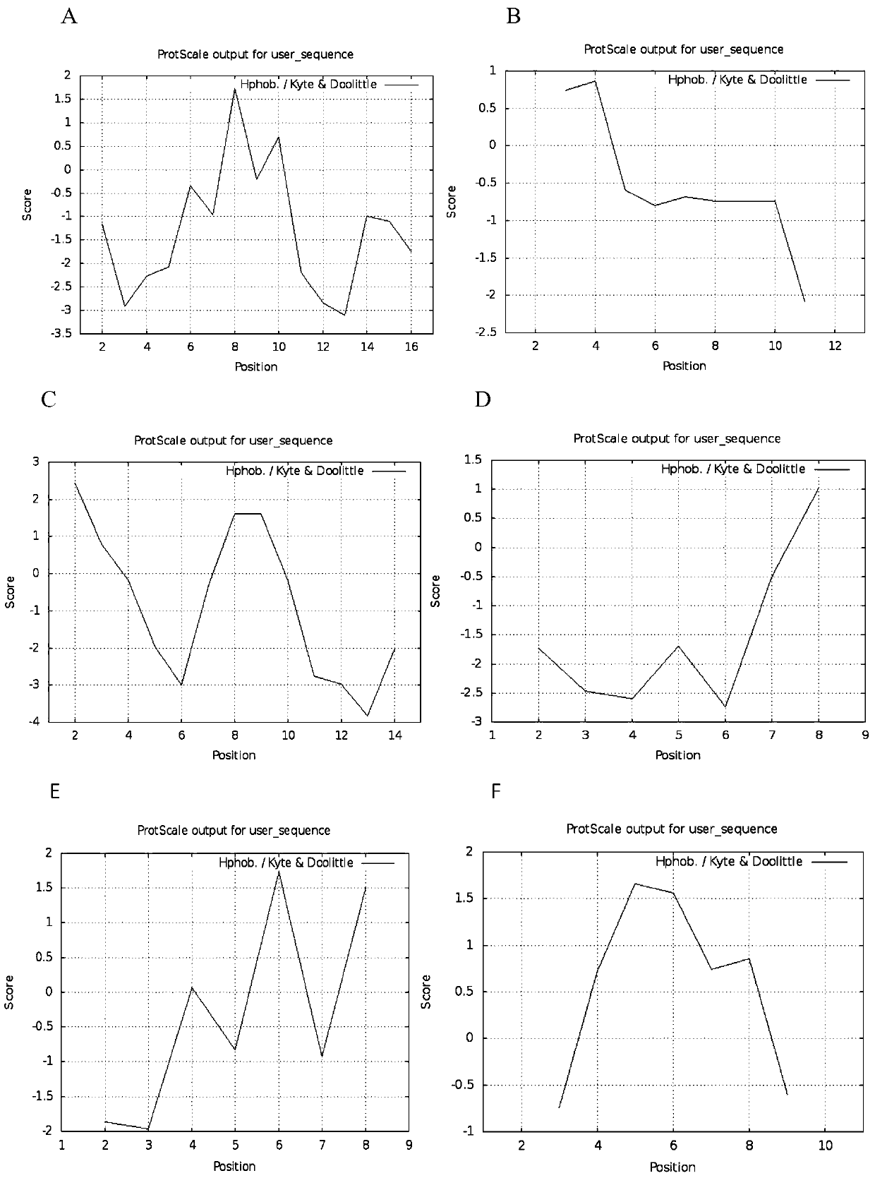
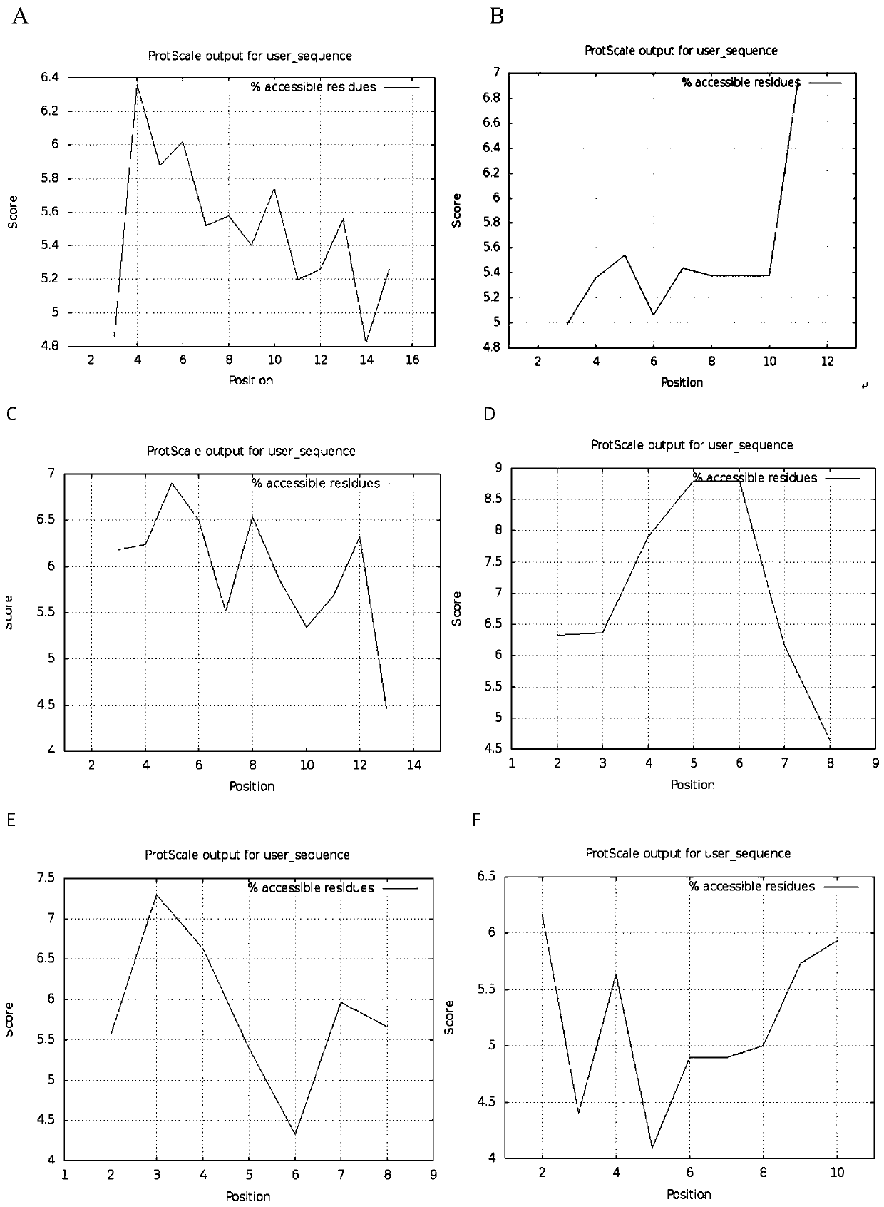
[0033] a using non-patent literature (Andreas Gloger, Danilo Ritz, Tim Fugmann, DarioNeri. Cancer Immunol Immunother, 2016, 65 (11): 1377-1393; Heyder T, Kohler M, Tarasova N K, et al. Approach for Identifying Human Leukocyte Antigen (HLA)-DRBound peptides from Scarce Clinical Samples.) The amino acid sequence described and synthesized and purified by GL Biochem Polypeptide Antigen 1 (IQRTPKIQVYSRHPAEN-Cys-KLH), Antigen 2 (YLLYYTEFTPTEK-Cys-KLH) and Antigen 3 (IQRTPKIQVYSRHPAEN -CYS-KLH-CYS-YLLYYTEFTPTEK) was prepared by ion chromatography. 1 mg of synthetic β2-MG polypeptide antigens 1, 2, 3 and natural β2-MG antigen were uniformly mixed with 1 ml of pH7.2 PBS solution and diluted to a concentration of 1 mg / ml antigen solution.

[0034] b Select 12 Balb / c mice with no significant difference in body weight (18-22g), week age (8w) and sex (female), and randomly divide them into three groups, namel...

example 2

[0037] The ELISA detection and identification of the monoclonal antibody of four kinds of antigens of anti-β2-MG of example 2

[0038] a The peptide detection antigen 1 (IQRTPKIQVYSRHPAEN-Cys-OVA), the peptide detection antigen 2 (YLLYYTEFTPTEK-Cys-OVA), the peptide detection antigen 3 (IQRTPKIQVYSRHPAEN-CYS-OVA-CYS- YLLYYTEFTPTEK) and β2-MG natural antigen solution were added to the small wells of the 96-well microtiter plate, placed overnight at 4°C, and the next day, the peptide detection antigen 1, peptide detection antigen 2, peptide detection antigen 3 and β2-MG The 96-well ELISA plate coated with the natural antigen solution was washed twice with washing solution (PBS buffer containing 0.05% Tween), and then blocked with a blocking solution (1% BSA) for two hours at room temperature. Pour off the blocking solution, pat dry on absorbent paper, paste the sealing paper and store at -20°C.

[0039] On the 10th-14th day after b fusion, the hybridoma cells obtained in Exampl...

example 3

[0043] Cloning of Example 3 Anti-β2-MG Natural Antigen (+) Monoclonal Antibody Hybridoma Cells

[0044] The positive hybridoma cell MCs obtained in Example 2 were diluted by the limiting dilution method, and positive single clones were selected for cloning culture. The specific method is as follows: the hybridoma cells against the natural antigen (+) obtained in Example 2 were counted respectively, and prepared into a single-cell suspension with a concentration of 1 cell / 200 μl through serial dilution, and 200 μl of the single-cell suspension was added to 96 wells In the small wells of the cell culture plate, culture in a 5% carbon dioxide incubator at 37°C. After culturing for about 9 days, select the supernatant of the monoclonal well for indirect ELISA detection, and set up a negative control (PBS) and a positive control (β2-MG antigen immune titer High mouse eye serum), select monoclonal hybridoma cells whose absorbance (OD) value is higher than the positive control well a...


PUM


Abstract

The invention discloses a preparation method of a beta2-microglobulin monoclonal antibody. The method comprises the following steps: firstly, screening out two polypeptide epitope sequences with relatively strong immunogenicity as a first antigen epitope and a second antigen epitope respectively; then respectively connecting one cysteine to each carbon-terminal amino acid of the first antigen epitope and the second antigen epitope, and coupling sulfydryl of the cysteine with primary amino of keyhole limpet hemocyanin through an SMCC coupling method so as to obtain an in-vitro synthesized beta2-MG double-epitope polypeptide antigen; then carrying out conventional immunization on the mouse by utilizing the double-epitope polypeptide antigen, and separating out splenic lymphocytes and myelomacells to carry out cell fusion; and finally, screening out positive monoclonal cells, and extracting monoclonal antibody through intraperitoneal injection and ascites recovery. By improving traditional single-epitope antigen immunization methods, the method optimizes the immunogenicity of the antigen, shortens the immunization period, and improves the serum titer in the immunization stage, thereby improving the obtaining efficiency of the positive monoclonal antibody against beta2-MG.

Description

technical field [0001] The invention relates to the field of biotechnology, in particular to a method for preparing a β2-microglobulin monoclonal antibody. Background technique [0002] β2 microglobulin, referred to as β2-MG, is a small molecular globulin secreted from lymphocytes, platelets and polymorphonuclear leukocytes. It is a single-chain linear polypeptide composed of 99 amino acids with a molecular weight of 11,800. β2-MG is a component of HLA-class I molecules. The concentration of β2-MG in normal human serum is in the range of 0.5-2.0mg / L. The only organ that excretes β2-MG out of the body is the kidney. When renal insufficiency occurs When β2-MG is excreted, the concentration of β2-MG in the patient's serum is 60 times that of normal people. When β2-MG is excessively deposited in the tissue, it is easy to induce β2-MG-related amyloidosis. Studies have found that the level of β2-MG in the serum is significantly negatively correlated with the glomerular filtration...

Claims


Application Information

Patent Timeline
Patent Type & Authority Applications(China)
IPC IPC(8): C07K16/28
CPCC07K16/2833C07K2317/34
Inventor 高健陈旭华汪渊谌敦华
Owner 上海钹乐诗生物技术有限公司
Learn More