Monoclonal antibody against filamentous virus GP protein and application of monoclonal antibody

A monoclonal antibody and antigen technology, applied in the direction of antiviral immunoglobulin, antiviral agent, application, etc., to achieve the effect of high homology and humanization

Active Publication Date: 2020-01-07
GUANGZHOU INST OF BIOMEDICINE & HEALTH CHINESE ACAD OF SCI
View PDF6 Cites 3 Cited by
  • Summary
  • Abstract
  • Description
  • Claims
  • Application Information

AI Technical Summary

Problems solved by technology

[0007] Aiming at the deficiencies of the prior art, the present invention provides a monoclonal antibody against filovirus GP protein and its application. The monoclonal antibody is prepared based on a macaque experimental technology platform, which solves the problem of the filovirus monoclonal antibody in practice. Diagnostic and therapeutic issues, providing new options for establishing filovirus detection, diagnosis, prevention and treatment

Method used


Image

  • Monoclonal antibody against filamentous virus GP protein and application of monoclonal antibody
  • Monoclonal antibody against filamentous virus GP protein and application of monoclonal antibody
  • Monoclonal antibody against filamentous virus GP protein and application of monoclonal antibody

Examples


Embodiment 2

[0080] Example 2 Marking and sorting of antigen-specific B cells

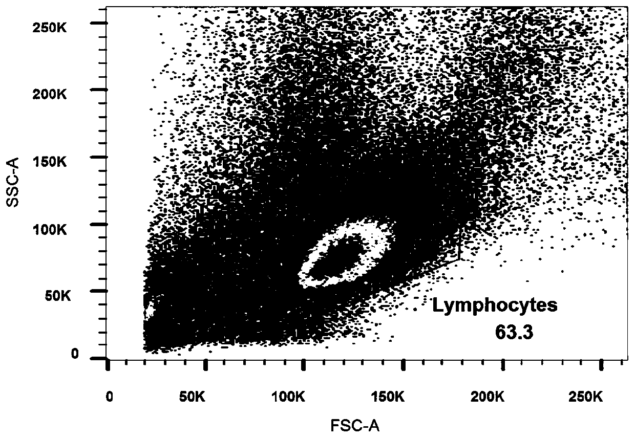
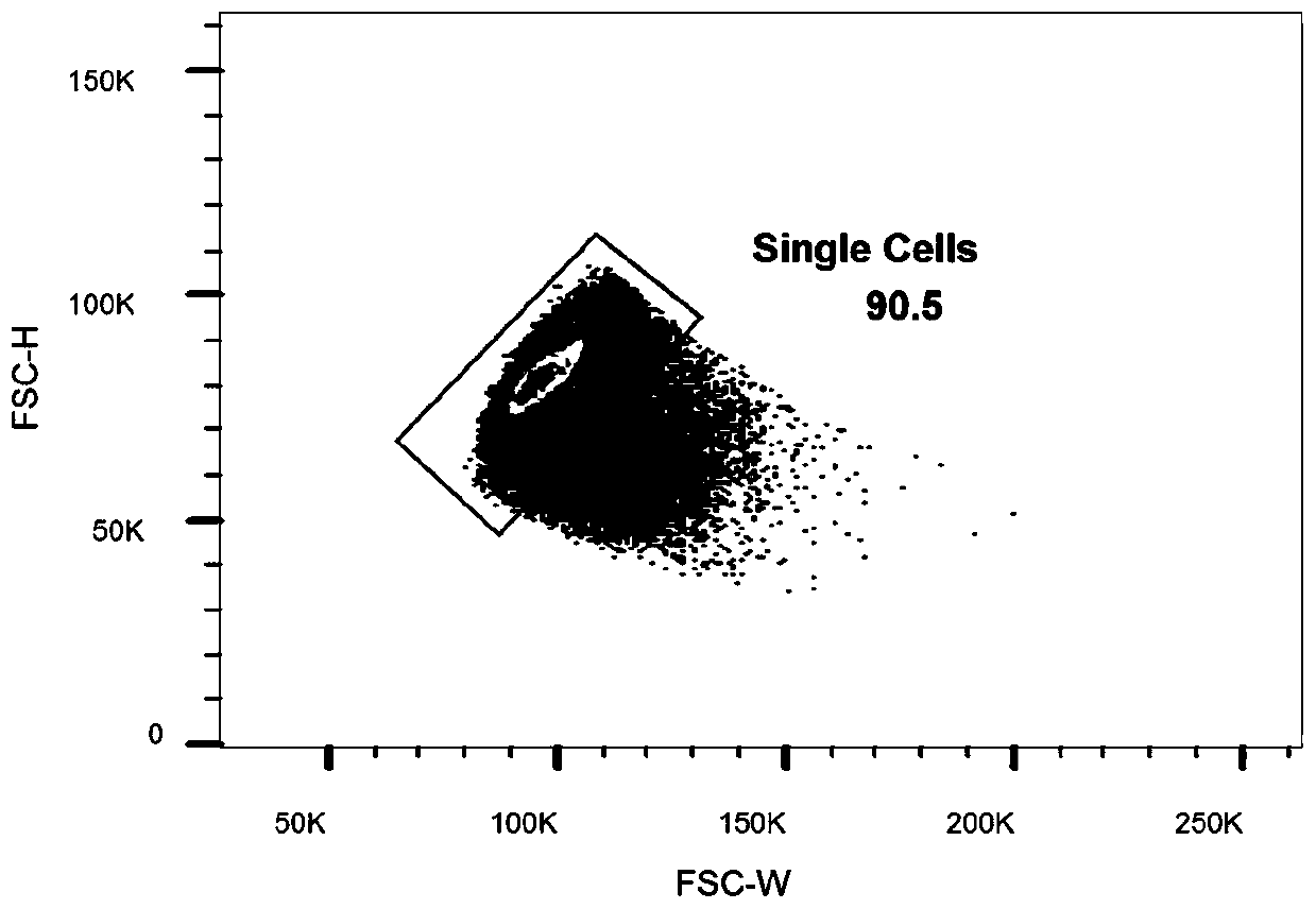
[0081] The obtained rhesus monkey PBMC was labeled with antibody labeling technology, CD3 antibody, CD20 antibody, CD27 antibody, IgG antibody and anti-histidine antibody, and a single GP protein-specific B cell was sorted by flow cytometry. Proceed as follows:

[0082] (1) Centrifuge the prepared PBMCs to discard the incubation supernatant, add 1 mL of PBS buffer, centrifuge at 400×g for 5 min, and discard the supernatant;

[0083] (2) First add 1 μL Aqua, incubate at 4°C for 20 minutes, then add 1 mL PBS, centrifuge at 400×g for 5 minutes, and discard the supernatant;

[0084] (3) Add the fluorescently labeled antibodies shown in Table 1, stain at 4°C for 30 minutes, add 1 mL of PBS after staining, centrifuge at 400×g for 5 minutes, and finally add 300 μL of PBS to resuspend, incubate at 4°C in a refrigerator, and perform sorting on the machine.

[0085] Table 1 Antibody labeling system

[0086]

[0087...

Embodiment 3

[0088] Example 3 Isolation of Antibody Variable Region Genes from Single B Cells Using RT-PCR

[0089]The reverse transcription kit produced by Promega was used to prepare the cell lysate, which was divided into 96-well plates, and the cells were centrifuged. After adding the reverse transcription reagent, they were reversed into cDNA. After the reverse transcription was completed, they were stored at -80°C;

[0090] Using rhesus monkey-specific primers to amplify the heavy and light chain variable region genes of the antibody through nested PCR using B cell cDNA as a template, the 50 μL system contains 5 μL cDNA, HotStarTaq Plus enzyme, dNTPs, and 0.5 μM specific primers, Carry out PCR amplification according to the following conditions: pre-denaturation at 94°C for 5min; 94°C for 30s, 55°C for 30s, 72°C for 50s, 35 cycles; 72°C for 7min; the obtained PCR product was identified by 1% agarose gel electrophoresis, the results image 3 shown.

[0091] The target fragments were ...

Embodiment 4

[0092] Embodiment 4 constructs the expression vector of monoclonal antibody

[0093] Use homologous recombination primers to add homologous recombination arms at the two ends of the antibody heavy chain variable region gene and the two ends of the light variable region gene, and use double enzymes to linearize the expression plasmid containing the human antibody heavy and light chain IgG1 constant regions Homologous recombination arm is generated; the variable region gene fragment added to the homologous recombination arm and the linearized plasmid are connected by homologous recombination to form a complete expression vector, in which the heavy chain of the monoclonal antibody is humanized The plasmid map is shown in Figure 4(A) and the light chain humanized plasmid map is shown in Figure 4(B). The recombinant product was transformed into TOP10 Escherichia coli competent to amplify the plasmid.


PUM


Abstract

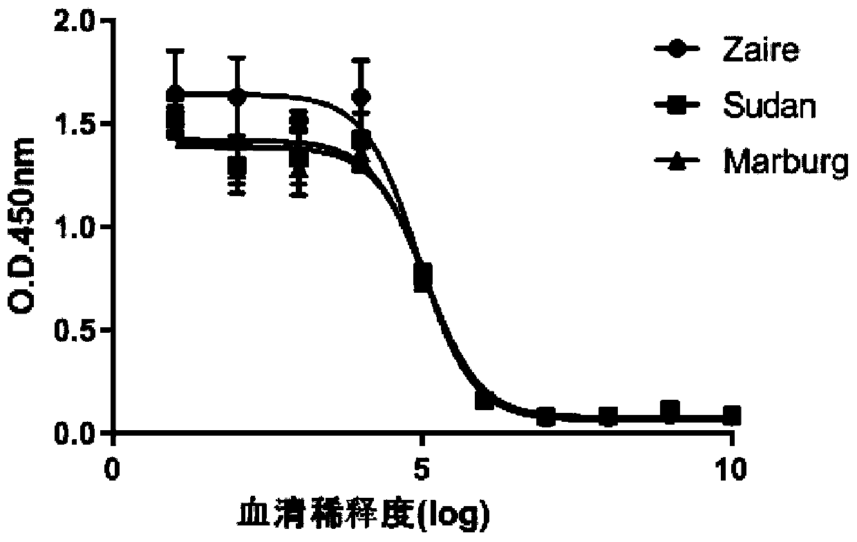
The present invention provides a monoclonal antibody against a filamentous virus GP protein and an application of the monoclonal antibody. A heavy chain variable region of an antigen-binding fragmentof the monoclonal antibody comprises an amino acid sequence shown as SEQ NO:7; and a light chain variable region of the antigen-binding fragment comprises an amino acid sequence shown as SEQ NO:8. Theantigen-binding fragment is obtained from an adult Chinese macaque monkey body, after humanization, the antigen-binding fragment has binding activity to Zaire-type GP protein, Sudan-type GP protein and Marburg virus GP protein of filamentous virus family. The obtained antibody has high homology with human genes, realizes humanization, is good in stability, has a relatively strong affinity for theGP proteins, can be potentially applied to antigen detection of filamentous viruses including Ebola viruses and Marburg viruses, detection and identification of clinical samples of filamentous virusinfection, development of filamentous virus inhibitors, etc.

Description

technical field [0001] The invention belongs to the field of biotechnology, and relates to a monoclonal antibody against filovirus GP protein and application thereof. Background technique [0002] Filoviruses are single-stranded negative-sense RNA viruses that are mainly divided into genera Ebolavirus, Marburgvirus and Cuevavirus. The Ebola virus genus includes five species: Zaire type (ZEBOV), Sudan type (SUDV), Reston type (RESTV), Tai Forest type (TAFV) and Bundibugyo type (BDBV). The Zaire, Sudan and Bundibugyo viruses have a fatality rate as high as 90%. The Marburg virus genus includes a species of Marburg virus (MARAV), which has a high fatality rate of 83-90%. In 2014, the Zaire virus broke out in West Africa, causing a total of 11,372 deaths as of 2016. As of March 10, 2019, the 10th round of Ebola hemorrhagic fever outbreak in Congo has caused 603 deaths. At present, there is no specific marketed therapeutic drug in the world, so the research on the diagnosis a...

Claims


Application Information

Patent Timeline
Patent Type & Authority Applications(China)
IPC IPC(8): C07K16/10C12N15/13A61P31/14
CPCC07K16/10A61P31/14C07K2317/565C07K2317/56C07K2317/76C07K2317/24C07K2317/92Y02A50/30
Inventor 陈凌王龙雨冯立强冯玉鹏
Owner GUANGZHOU INST OF BIOMEDICINE & HEALTH CHINESE ACAD OF SCI
Why Eureka
Try Eureka