A kind of preparation method and device of ultrafine atomic force microscope metal probe

A technology of atomic force microscope and metal probe, which is applied in the direction of measuring device, scanning probe technology, scanning probe microscopy, etc., can solve the problems of large radius of curvature of the tip, easy to break, low reflectivity, etc., and achieve stability and Good electrical conductivity, not easy to fall off or wear, high preparation success rate

Active Publication Date: 2020-08-18
ZHEJIANG UNIV
View PDF6 Cites 0 Cited by
  • Summary
  • Abstract
  • Description
  • Claims
  • Application Information

AI Technical Summary

Problems solved by technology

However, silicon and silicon nitride materials have low reflectivity in the visible and near-infrared, and it is usually necessary to coat the cantilever with a metal layer to improve the reflectivity, which makes the radius of curvature of the tip larger and it is difficult to directly observe the bottom of the atomic-level structure. Case
Moreover, the shape of such traditional AFM probes is generally pyramidal or conical, with a large diameter, brittle texture, and easy to break
Carbon nanotubes have the advantages of small diameter and good flexibility, but the current main methods for preparing carbon nanotube AFM probes (including growth method, pickup method, electrophoresis method and mechanical manipulation method, etc.) cannot control the probe angle and long diameter. Ratio, it is difficult to obtain a single high-quality probe

Method used


Image

  • A kind of preparation method and device of ultrafine atomic force microscope metal probe
  • A kind of preparation method and device of ultrafine atomic force microscope metal probe
  • A kind of preparation method and device of ultrafine atomic force microscope metal probe

Examples


preparation example Construction

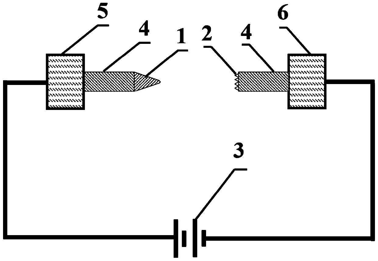
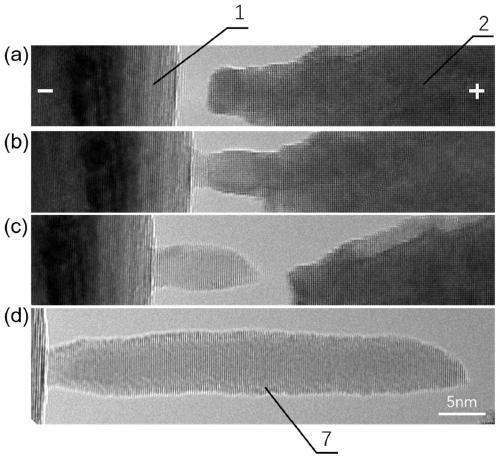
[0058] 4) Preparation of metal probes for atomic force microscopy: A certain amount of voltage is applied to both ends of the electrical-mechanical mono-tilt sample rod. - The movable end of the mechanical mono-tilt sample rod moves the movable end of the metal needle tip 1, so that the gap between the metal needle tip 1 and the metal fracture end 2 is close to about 2nm, and the close parts of both ends are connected to each other under the action of electromigration, A single crystal metal nanowire is formed, and then the movable end is slowly moved backwards at a uniform speed, and finally the two ends are separated to obtain a single ultrafine atomic force microscope metal probe 7 with a certain length;

[0059] 5) Repeat the above step 4) repeatedly to make the atomic force microscope metal probe grow stably along the axial direction, and obtain an atomic force microscope metal probe with an ideal length and shape through multiple adjustments, that is, an atomic force micr...

Embodiment 1

[0067] Such as image 3 and Figure 5 shown.

[0068] (1) Physical shearing to prepare the metal fracture end 2

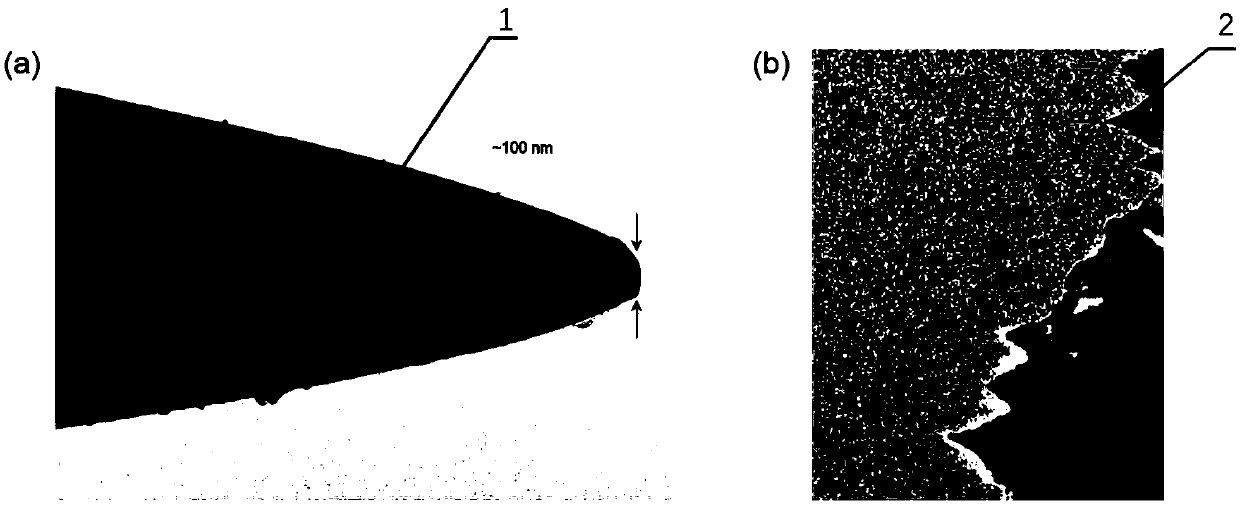
[0069] Sonicate a tungsten metal wire with a diameter of 0.25 mm in ethanol solution for 10 minutes, flatten it with a vise, and cut it with scissors. At this time, there will be a large number of nanometer-sized small protrusions on the front end of the wire to form a nanoscale rough surface, that is, At the metal fracture end 2, a large number of small protrusions of nanometer size exist at the front end, with different shapes and orientations. Fix the root of the metal fracture end 2 to the fixed end, and the flattened and cut end of the metal fracture end 2 faces the movable end.

[0070] (2) Electrochemical corrosion of tungsten wire

[0071] The specific implementation adopts tungsten metal wire, which is ultrasonically cleaned in ethanol solution before use to remove surface dirt. Configure 1mol / L sodium hydroxide solution, combined with an electrochemi...

Embodiment 2

[0081] The difference from Example 1 is that the growth direction is different, the metal needle tip 1 is connected to the negative pole of the power supply 3 , the metal fracture end 2 is connected to the positive pole of the power supply 3 , and the others are the same as in Example 1. The implementation process is shown as Figure 4 and Figure 6 As shown, the metal atom 8 is to the right.


PUM

PropertyMeasurementUnit
diameteraaaaaaaaaa
diameteraaaaaaaaaa
diameteraaaaaaaaaa

Abstract

The invention discloses a method and device for preparing a superfine atomic force microscope metal probe. A homogeneous metal fracture end and a metal probe tip are respectively prepared, the metal fracture end is arranged at the fixed end of an electrical-mechanical sample rod, the metal probe tip is loaded on the movable end of the electrical-mechanical sample rod, and the two ends are oppositely arranged without contact. In a transmission electron microscope, the metal probe tip aligns with and is close to a fracture sample by piezoelectric ceramic drive; and a constant bias voltage is applied two ends at a critical contact point, and the preparation of the superfine atomic force microscope metal probe is realized by utilizing the electro-atomic migration effect. The preparation methodof the atomic force microscope metal probe is high in success rate, the growth direction and the length of a finished product are controllable, the diameter of the tip is less than 5nm, and the stability and the conductivity are good.

Description

technical field [0001] The invention relates to the technical field of micro-nano manufacturing, in particular to a method and device for in-situ preparation of a controllable ultrafine atomic force microscope single metal probe. Background technique [0002] Atomic Force Microscope (AFM) is a micron-scale precision mechanical measuring instrument invented by Binning et al. in 1986. The AFM probe is one of the key components to achieve precise measurement. Constructed with a needle tip, where the microcantilever is extremely sensitive to weak forces. When the sample is image-scanned, the probe and the sample surface are close to each other, and the interaction force between the atoms at the tip of the probe and the surface atoms of the sample increases rapidly as the distance between them decreases, resulting in a small displacement of the cantilever beam. This small displacement is detected and used as feedback to keep the force constant to obtain the position change of th...

Claims


Application Information

Patent Timeline
Patent Type & Authority Patents(China)
IPC IPC(8): G01Q60/40
CPCG01Q60/40
Inventor 王江伟赵治宇韦华
Owner ZHEJIANG UNIV
Why Eureka
Try Eureka