Wild edible fungus tissue separation method

A technology for tissue separation and fungus, applied in the direction of microorganism-based methods, biochemical equipment and methods, and separation of microorganisms, can solve the problems of lack of standardized operation of fungus and efficient drying methods, high pollution rate, etc., to save time and reduce pollution efficiency, efficient operation

Inactive Publication Date: 2020-03-10
JILIN AGRICULTURAL UNIV
View PDF5 Cites 0 Cited by
  • Summary
  • Abstract
  • Description
  • Claims
  • Application Information

AI Technical Summary

Problems solved by technology

[0006] The purpose of the present invention is to provide a wild fungus tissue separation method to solve the problems of high pollution rate, lack of standardized operation and efficient drying method in the process from collection to tissue separation of fungus in the existing wild fungus tissue separation method

Method used


Image

  • Wild edible fungus tissue separation method
  • Wild edible fungus tissue separation method

Examples


Embodiment 1

[0017] Embodiment 1 A kind of wild fungus tissue separation method

[0018] 1) Collect dried ears: Collect complete, mildew-free, and insect-free fungus, gently wipe off surface debris, put them in a hot air drying box for drying, set the drying temperature at 65°C, and collect dry ears after drying for 4-8 hours. Ear;

[0019] 2) Tissue separation: Use sterile forceps to remove the thinner tissue on the edge of the dry ear, and then take 0.3-0.6cm 2 Small and large tissues, placed in the prepared ceftriaxone sodium alcohol solution, soaked for 3-5min, taken out with tweezers, quickly passed through the flame twice, and transferred to a plate culture dish filled with PDA medium;

[0020] 3) Separation culture: place the petri dish in a constant temperature incubator at 25°C and incubate in the dark until the mycelium grows to the medium, pick the tip of the mycelium and inoculate it on the PDA medium, and the strain can be preserved after it is full;

[0021] Described ceftr...

Embodiment 2

[0023] Example 2 Tissue Separation Example of Fresh Fruiting Body of Auricularia auriculae

[0024] 1. Collection and drying of fungus

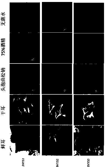
[0025] Record the habitat and tree species of 3 fresh fungus collected in Jilin Agricultural University, and select complete, mildew-free, and insect-free fungus to gently wipe off the surface debris. They are numbered JNYS1, JNYS2, and JNYS3 respectively. After recording, put it into a ziplock bag; put the collected specimens into a hot air drying box for drying, set the drying temperature at 60°C, and collect them after drying for 5 hours;

[0026] 2. Preparation of disinfectant

[0027] Take out 5ml of sterile water, pour it into the ceftriaxone sodium medicine bottle, beat it repeatedly until it is completely dissolved, take out 3ml of the medicine solution and pour it into a sterile glass plate filled with 75% alcohol and set the volume to 40ml, and prepare the ceftriaxone sodium alcohol solution. Beat and mix repeatedly so that the co...


PUM


Abstract

The invention discloses a wild edible fungus tissue separation method. The method comprises the following steps: 1) collecting edible fungus, removing impurities, and drying the edible fungus at 55-65DEG C to obtain dried edible fungus, (2) taking 0.3-0.6 cm<2> of tissues of the dried edible fungus, and soaking the tissues in a ceftriaxone sodium alcohol solution for 3-5 minutes, and (3) culturing the tissues in a dark place at 22-28 DEG C for 3-4 days, and inoculating a PDA (potato dextrose agar) culture medium with the picked hypha tips. The method has the advantages that 1) compared with low-temperature drying (at 30 DEG C or below) and natural drying, time is saved, the pollution rate caused by a non-laboratory environment is reduced, and a survival rate of collected wild edible fungus resources is increased, 2) only a single antibiotic alcohol solution is used for soaking treatment in the operation process, mercury bichloride and other drugs are not used, and operation is harmless and efficient, 3) according to the tissue separation method, the hypha germination phenomenon can be observed on the third day of inoculation, and the pure strain can be obtained by purification again, and 4) the method is suitable for black fungus, auricularia polytricha and other edible and medicinal macro-fungi with edible and medicinal values which are widely cultivated at present.

Description

technical field [0001] The invention belongs to the technical field of strain separation, and in particular relates to a method for separating wild fungus tissue. Background technique [0002] Wild germplasm resources can be used as excellent breeding materials to screen highly resistant strains; to solve the problem of degeneration of main plant varieties; to enrich germplasm resource banks and protect the diversity of germplasm resources; to enrich genetic resources and genetic diversity research. The excavation of wild germplasm resources of fungus is of great significance to the development of edible fungus industry. [0003] Fungus edible fungus has a long history of edible and medicinal use, and it is recorded in ancient books such as "Shen Nong's Materia Medica", "Bielu of Famous Doctors", "Qi Min Yao Shu", "Fungus Spectrum" and so on. At present, the output of black fungus and fungus in my country ranks second, second only to shiitake mushrooms. They have a wide cul...

Claims


Application Information

Patent Timeline
Patent Type & Authority Applications(China)
IPC IPC(8): C12N1/02C12N1/14C12R1/645
CPCC12N1/02C12N1/14
Inventor 宋冰陈艳琦李玉吕志文吕艳聪刘柱衫付永平
Owner JILIN AGRICULTURAL UNIV
Why Eureka
Try Eureka