Cell stripping method
A technology of cells and cell fluids, which is applied in the field of medicine to achieve the effect of improving the stripping efficiency, simplifying the experimental process, and improving the efficiency of stripping and purification
- Summary
- Abstract
- Description
- Claims
- Application Information
AI Technical Summary
Problems solved by technology
Method used
Image
Examples
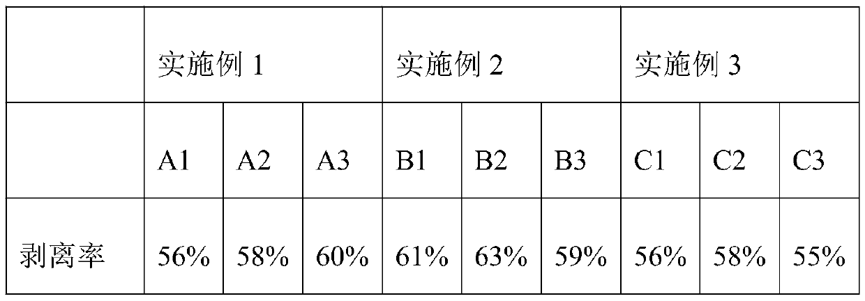
Embodiment 1
[0022] S1. Put 40g of nylon wool into a beaker, add 80ml of distilled water, cover the beaker with aluminum foil and boil for 15 minutes; then cool to room temperature, pour it into a funnel with filter paper, filter out the water, and put the nylon wool again In the beaker, repeat the above steps 5 times;
[0023] S2. Spread the nylon wool in a tray covered with gauze, incubator at 30℃ and dry for 30h;
[0024] S3. Take a 50ml syringe, and configure a rubber tube with a collet at the mouth of the syringe, put nylon wool into the syringe, then wrap the syringe with tin foil, and put it into a sterilization box for sterilization.
[0025] S4. Fix the syringe, fill the syringe with a cell culture solution at 35°C, let it stand for 30 minutes, then open the chuck, and release the cell culture solution, and repeat the above steps twice.
[0026] S5. Dilute the cell liquid to be stripped to 4.9×10 with the culture medium at 35°C 7 cells / ml
[0027] S6. Pour the cell liquid in S5...
Embodiment 2
[0030] S1. Put 60g of nylon wool into a beaker, add 120ml of distilled water, cover the beaker with aluminum foil and boil for 18 minutes; then cool to room temperature, pour it into a funnel with filter paper, filter out the water, and put the nylon wool again In the beaker, repeat the above steps 6 times;
[0031] S2. Spread the nylon wool in a tray covered with gauze, incubate at 35°C and dry for 35h, cover the top of the tray with aluminum foil;
[0032] S3. Take a 50ml syringe, and configure a rubber tube with a collet at the mouth of the syringe, put nylon wool into the syringe, then wrap the syringe with tin foil, and put it into a sterilization box for sterilization.
[0033] S4. Fix the syringe, fill the syringe with a cell culture solution at 38°C, let it stand for 30 minutes, then open the chuck, and let off the cell culture solution, and repeat the above steps 3 times.
[0034] S5. Dilute the cell liquid to be stripped to 5×10 with the culture medium at 38°C 7 ce...
Embodiment 3
[0038] S1. Put 80g of nylon wool into a beaker, add 160ml of distilled water, cover the beaker with aluminum foil and boil for 20min; then cool to room temperature, pour it into a funnel with filter paper, filter out the water, and put the nylon wool again In the beaker, repeat the above steps 7 times;
[0039] S2. Spread the nylon wool in a tray covered with gauze, incubator at 45℃ and dry for 40h;
[0040] S3. Take a 50ml syringe, and configure a rubber tube with a collet at the mouth of the syringe, put nylon wool into the syringe, then wrap the syringe with tin foil, and put it into a sterilization box for sterilization.
[0041] S4. Fix the syringe, fill the syringe with cell culture solution at 40°C, let it stand for 30 minutes, then open the chuck, and let off the cell culture solution, and repeat the above steps 4 times.
[0042] S5. Dilute the cell liquid to be stripped with a culture medium at 40°C to 5.1×10 7 cells / ml
[0043] S6. Pour the cell liquid in S5 into ...
PUM
 Abstract
Description
Claims
Application Information
 - R&D
- Intellectual Property
- Life Sciences
- Materials
- Tech Scout
- Unparalleled Data Quality
- Higher Quality Content
- 60% Fewer Hallucinations
Browse by: Latest US Patents, China's latest patents, Technical Efficacy Thesaurus, Application Domain, Technology Topic, Popular Technical Reports.
© 2025 PatSnap. All rights reserved.Legal|Privacy policy|Modern Slavery Act Transparency Statement|Sitemap|About US| Contact US: help@patsnap.com

