Eureka AIR delivers breakthrough ideas for toughest innovation challenges, trusted by R&D personnel around the world.

Application of fluorescence in-situ hybridization on bone marrow smears in multiple myeloma

A technique of fluorescence in situ hybridization and multiple myeloma, which is applied in the determination/testing of microorganisms, biochemical equipment and methods, etc., can solve the problems of low false negative rate, increased cost, and low proportion of plasma cells, and achieve simple operation Effect

Inactive Publication Date: 2020-04-14
WUHAN UNIV
View PDF6 Cites 1 Cited by
  • Summary
  • Abstract
  • Description
  • Claims
  • Application Information

AI Technical Summary

Problems solved by technology

[0006] Usually, we use heparin anticoagulated bone marrow specimens to harvest nucleated cells for FISH probe detection. Since multiple myeloma cells are focally distributed in the bone marrow, when the aspirated bone marrow exceeds 0.2 ml, it is easily diluted by peripheral blood. Currently, myeloma Most cell enrichment detection methods are CD138 immunomagnetic bead sorting combined with FISH and cytoplasmic κ / λ light chain immunofluorescence combined with FISH (cIg-FISH) to identify plasma cells, and then perform FISH detection, which has a low false negative rate and is suitable for plasma cells Low proportion, MRD monitoring, but the manual operation steps of these two methods are complicated, time-consuming and costly

Method used


Image

  • Application of fluorescence in-situ hybridization on bone marrow smears in multiple myeloma
  • Application of fluorescence in-situ hybridization on bone marrow smears in multiple myeloma
  • Application of fluorescence in-situ hybridization on bone marrow smears in multiple myeloma

Examples


Embodiment 1

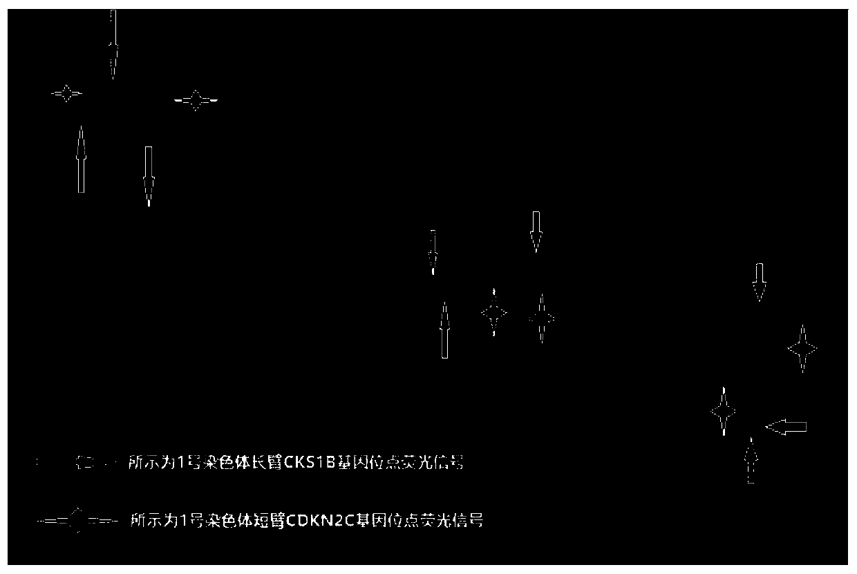
[0032] In a patient with multiple myeloma, the ilium bone marrow smear cell morphology of myeloma cells was 57%, the cell body size was different, the cytoplasm was increased, the nucleus was enlarged, the cytoplasm was light blue, and the nucleus was round or oval , visible giant, dual / multinucleated tumor cells (eg figure 1 shown). At the same time, the first tube of heparin-anticoagulated bone marrow aspirated at the same time was detected by flow cytometry, and only a group of abnormal monoclonal plasma cells accounted for 3.7% of bone marrow nucleated cells were found. The proportion of myeloma cells in the subsequently aspirated heparin-anticoagulated bone marrow was even lower. CD138 magnetic bead sorting requires more than 20 mL of bone marrow to obtain a sufficient amount of myeloma cells for FISH detection. In this embodiment, a bone marrow smear of less than 0.2 mL is selected for fluorescence in situ hybridization detection, and the specific operations are as foll...

Embodiment 2

[0058] In a patient with multiple myeloma, myeloma cells accounted for 10% of the iliac bone marrow smear cells, with large cell bodies, increased cytoplasm, enlarged nuclei, blue cytoplasm, round and oval nuclei (such as Figure 4 shown). At the same time, the first tube of heparin-anticoagulated bone marrow aspirated at the same time was detected by flow cytometry immunotype, only a group of monoclonal abnormal plasma cells accounted for 2.3% of bone marrow nucleated cells, and the proportion of myeloma cells in the subsequently aspirated heparin-anticoagulated bone marrow was even lower. CD138 magnetic bead sorting and aspirating more than 20mL of bone marrow still cannot obtain enough myeloma cells for FISH detection. In this embodiment, a bone marrow smear of less than 0.2 mL is selected for fluorescence in situ hybridization detection, and the specific operations are as follows:

[0059] 1. Preparation of Bone Marrow Smear

[0060] 1.1 Preparation of slides: quickly pl...

Embodiment 3

[0084] In a patient with multiple myeloma, myeloma cells accounted for only 3% of the iliac bone marrow smear cells, with small cell bodies, varying amounts of cytoplasm, light blue cytoplasm, and round or oval nuclei (such as Figure 8 shown). At the same time, the first tube of heparin-anticoagulated bone marrow aspirated at the same time was detected by flow cytometry immunotype, only a group of monoclonal abnormal plasma cells accounted for 0.2% of bone marrow nucleated cells, and the proportion of myeloma cells in the subsequent heparin-anticoagulated bone marrow was even lower. Sorting with CD138 magnetic beads cannot obtain enough myeloma cells for FISH detection. In this embodiment, bone marrow smear fluorescent in situ hybridization technology is selected for detection, and the specific operations are as follows:

[0085] 1. Preparation of Bone Marrow Smear

[0086] 1.1 Preparation of slides: quickly place 0.2mL of bone marrow fluid aspirated for the first time on a c...


PUM


Abstract

The invention belongs to the field of tumor molecular diagnosis, and particularly relates to application of fluorescence in-situ hybridization on bone marrow smears in multiple myeloma. The application specifically comprises the following steps: making a bone marrow fluid into bone marrow smears for Wright's staining, and selecting an area with more myeloma cells and good dispersion as an FISH (fluorescence in-situ hybridization) area; covering the FISH area with a fresh stationary liquid to fade the FISH area and fix the cells, washing the bone marrow smears in a 2*SSC solution of 56 DEG C for 10 min, rapidly rinsing the bone marrow smears in deionized water once, and then putting the bone marrow smears into gradient ethanol for dehydration and air-drying; adding a FISH probe into the FISH area for hybridization, dyeing by using a DAPI counterstaining agent, and carrying out microscopic examination on a fluorescence hybridization signal. The bone marrow smear FISH detection method isespecially suitable for the condition that the proportion of monoclonal abnormal plasma cells in flow immunotyping is lower than 20%, and the proportion of myeloma cells in bone marrow smear Wright'sstaining morphology is higher than 20%.

Description

technical field [0001] The invention belongs to the field of tumor molecular diagnosis, and in particular relates to the application of a bone marrow smear fluorescence in situ hybridization technique in multiple myeloma. Background technique [0002] Multiple myeloma (MM, multiple myeloma) is a malignant plasma cell proliferative disease with strong biological heterogeneity, large individual differences in clinical efficacy, and survival ranging from months to decades. Cytogenetic and molecular genetic characteristics are important prognostic indicators for MM. The prognostic stratification system and individualized treatment based on genetic characteristics have become the development trend of MM clinical research. [0003] Fluorescence in situ hybridization (FISH, fluorescence in situ hybridization) technology uses DNA denaturation to untangle the double strands to become single strands, which can form stable heteroduplexes with complementary DNA strands after annealing a...

Claims


Application Information

Patent Timeline
IPC IPC(8): C12Q1/6841
CPCC12Q1/6841C12Q2563/107
Inventor 喻亚兰周芙玲童西琴刘莉沈辉吴三云罗萍
Owner WUHAN UNIV
Why Eureka
Eureka Blog
Learn More