Triple fluorescent quantitative PCR detection material and kit for distinguishing African swine fever virus wild strains from CD2V and/or 360-505R gene deleted strains

A fluorescence quantitative and gene deletion technology, applied in the field of virus strain identification, can solve the problem of inability to distinguish the infection of wild virus strains with gene deletion strains, and achieve the effects of saving detection cost and time, high sensitivity, and convenient clinical application and operation.

Active Publication Date: 2020-04-28
SOUTH CHINA AGRI UNIV +1
View PDF6 Cites 13 Cited by
  • Summary
  • Abstract
  • Description
  • Claims
  • Application Information

AI Technical Summary

Problems solved by technology

However, these methods mainly detect the conserved capsid protein gene P72, and cannot distinguish between wild strains and gene deletion strains.

Method used


Image

  • Triple fluorescent quantitative PCR detection material and kit for distinguishing African swine fever virus wild strains from CD2V and/or 360-505R gene deleted strains
  • Triple fluorescent quantitative PCR detection material and kit for distinguishing African swine fever virus wild strains from CD2V and/or 360-505R gene deleted strains
  • Triple fluorescent quantitative PCR detection material and kit for distinguishing African swine fever virus wild strains from CD2V and/or 360-505R gene deleted strains

Examples


Embodiment 1

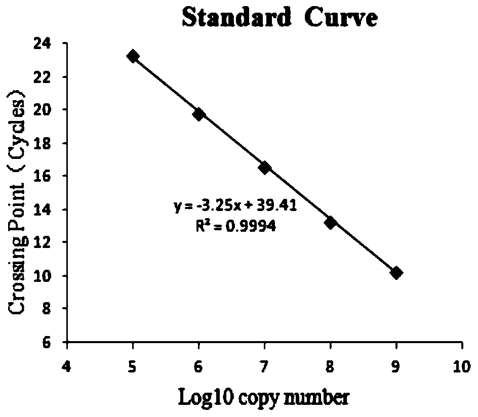
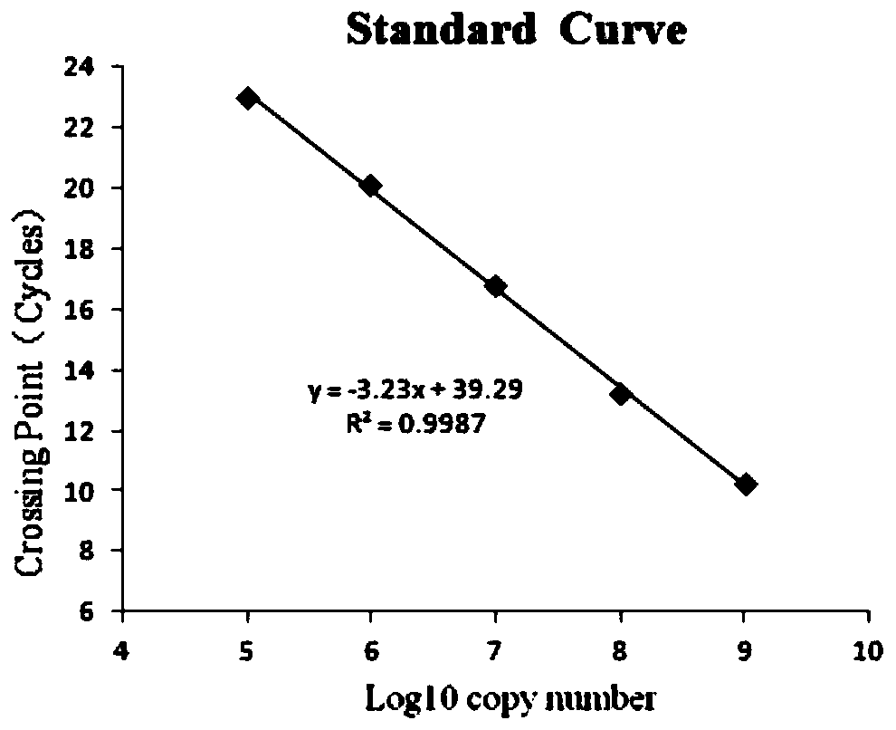
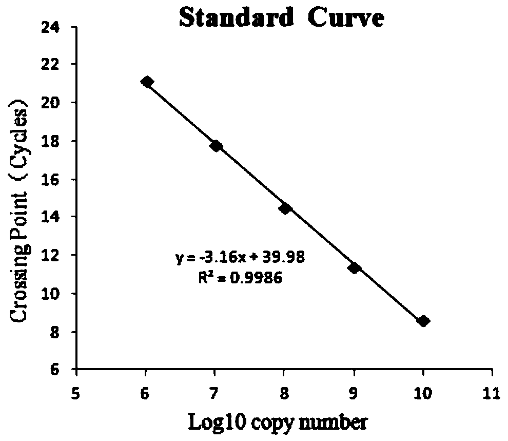
[0066] Embodiment 1 Primer and probe design

[0067] The inventor compared the P72, CD2V and 360-505R genes of African swine fever virus (ASFV) published in the GenBanK database of NCBI (National Center for Bioinformatics of the United States) in a large number of highly conserved and specific regions, and designed ASFV P72, CD2V and 360 The specific primers and probes for the -505R gene correspond to the three groups A, B, and C respectively. The specific sequences are:

[0068] Group A:

[0069] Upstream primer ASFV-P72-F:

[0070] TTTAATGAGAACGTGAACCTTGCT (SEQ ID NO: 1);

[0071] Downstream primer ASFV-P72-R:

[0072] ATGGTTTAAAGCGTATATTGCGTCT (SEQ ID NO: 2);

[0073] Specific fluorescent probe ASFV-P72-P:

[0074] Fluorophore - TCCCTCAGTATCCATTCCCTTCGGCGAG - Quencher (SEQ ID NO: 3);

[0075] Group B:

[0076] Upstream primer ASFV-CD2V-F:

[0077] CCTAAGCCTTACAGTCGTTATCAG (SEQ ID NO: 4);

[0078] Downstream primer ASFV-CD2V-R:

[0079] ACATGGTTTAGGTGGAGGACAT (SEQ ...

Embodiment 2 3

[0090] Embodiment 2 Triple fluorescent quantitative PCR detection method

[0091] Materials and Methods

[0092] 1.1 Primers in Example 1

[0093] 1.2 Sample DNA extraction

[0094] There are no special requirements for DNA extraction, which can be extracted according to conventional methods or DNA extraction kits. The extracted DNA was stored at -20°C for later use or immediately used for PCR amplification.

[0095] 1.3 Positive plasmid

[0096] Connect the PCR amplification products of three sets of primers A, B, and C to the pJET1.2 cloning vector, transform Escherichia coli competent cells DH5α, spread on LB medium plates containing 100mg / L ampicillin, and culture at 37°C After 12-16 hours, after picking bacteria, screening and sequencing identification, the plasmids were extracted after expanding the culture of positive bacteria, and the positive plasmids were named pJET1.2-P72(qPCR), pJET1.2-CD2V(qPCR), pJET1.2-360 -505R (qPCR).

[0097] 1.4 Triple fluorescent quan...

Embodiment 3

[0118] Embodiment 3 sensitivity test

[0119] Dilute the positive sample concentration to 1×10 -3 ng / μL, 1×10 -2 ng / μL, 1×10 -1 ng / μL, 1×10 0 ng / μL, 1×10 1 ng / μL.

[0120] The above-mentioned different template concentrations were detected according to the triple fluorescent quantitative PCR detection method established in 1.4 of Example 2. The results are attached Figure 5 shown. From attached Figure 5 As can be seen in the 1×10 -3 The sample at the concentration of ng / μL still had a good amplification curve, indicating that the sensitivity of the method reached 1×10 -3 ng / μL.


PUM

PropertyMeasurementUnit
diameteraaaaaaaaaa

Abstract

The invention discloses a triple fluorescent quantitative PCR detection material and kit for distinguishing African swine fever virus wild strains from CD2V and/or 360-505R gene deleted strains. The detection material comprises primers and probes, and the nucleotide sequences are shown as SEQ ID NO: 1-9. According to the invention, triple fluorescent quantitative PCR amplification is carried out on African swine fever virus P72, CD2V and 360-505R genes by utilizing three groups of primer probes, so that the detection cost and the detection time for identifying different genes are reduced; whether gene deletion exists in the strain or not can be distinguished; the detection material and kit have high specificity and sensitivity; and for traditional qPCR enabling only one gene to be amplified each time, the provided mode has the following advantages: three genes are amplified at the same time in each PCR reaction, and the cost is reduced by about 2/3.

Description

technical field [0001] The invention relates to the technical field of virus strain identification, and more specifically relates to a triple fluorescent quantitative PCR detection material and a kit for distinguishing ASFV wild strains from double gene deletion strains. Background technique [0002] African swine fever (ASF) is caused by African swine fever virus (ASFV), which can cause acute hemorrhagic fever, resulting in a large number of morbidity and mortality events (mortality close to 100%) in pigs. The World Organization for Animal Health (OIE) listed it as a must-report animal disease, and my country listed it as a first-class animal disease. Since the first case of African swine fever broke out in my country in 2018, African swine fever has caused major losses to my country's pig industry and seriously affected the healthy development of my country's pig industry. African swine fever has spread to many countries in Asia. The impact of African swine fever on my c...

Claims


Application Information

Patent Timeline
Patent Type & Authority Applications(China)
IPC IPC(8): C12Q1/70C12Q1/6851C12N15/11C12R1/93
CPCC12Q1/701C12Q1/6851C12Q2600/16C12Q2531/113C12Q2537/143C12Q2561/101C12Q2545/114Y02A50/30
Inventor 沈永义张旭陈瑞爱沈雪娟李延鹏张文炎
Owner SOUTH CHINA AGRI UNIV
Why Eureka
Try Eureka