Medical image segmentation method based on deep learning

A technology of medical imaging and deep learning, applied in the fields of medical image processing and computer vision, can solve the problems of redundant computing resources and model parameters, unbalanced medical image data, small perceptual area, etc., and achieve feature reuse and optimal segmentation effect, the effect of suppressing the response

Active Publication Date: 2020-05-12
UNIV OF ELECTRONICS SCI & TECH OF CHINA +1
View PDF10 Cites 38 Cited by
  • Summary
  • Abstract
  • Description
  • Claims
  • Application Information

AI Technical Summary

Problems solved by technology

However, the traditional CNN cannot reasonably propagate the low-level features to the high-level, so the U-Net algorithm is further proposed, which fuses low-dimensional and high-dimensional features through skip connections, and has a good segmentation effect
[0004] Most of the current medical image segmentation algorithms are based on U-Net (U-Net Baseline), but due to the problems of data imbalance, large differences between training samples, and small perceptual regions (ROI) in medical images, the accuracy and It is difficult to balance the recall rate, insufficient feature extraction, redundant computing resources and model parameters, and the segmentation effect is not obvious, so more accurate segmentation of ultrasound images is still a problem that needs to be solved urgently

Method used


Image

  • Medical image segmentation method based on deep learning
  • Medical image segmentation method based on deep learning
  • Medical image segmentation method based on deep learning

Examples


Embodiment Construction

[0031] Below in conjunction with accompanying drawing and emulation the present invention is described in detail:

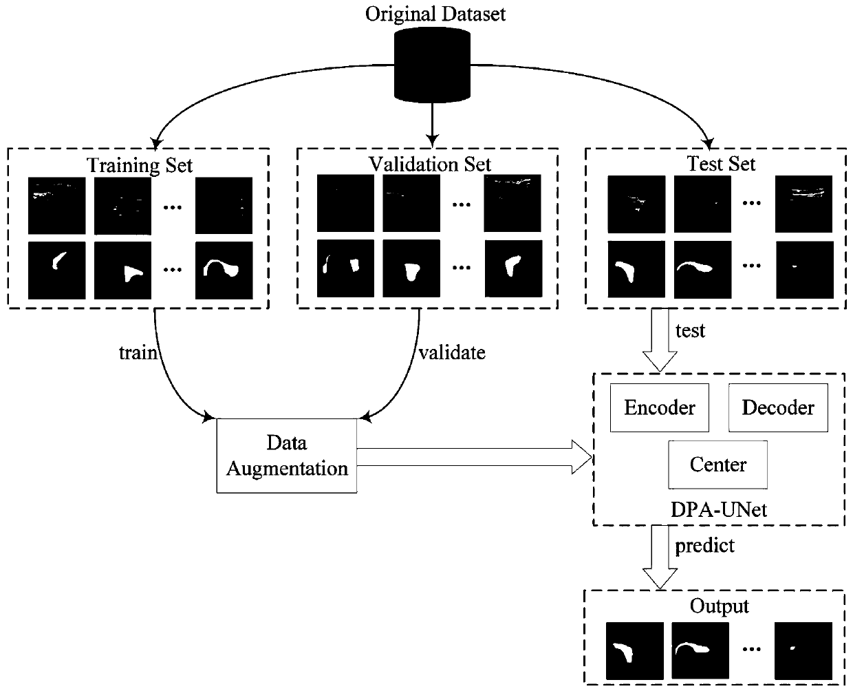
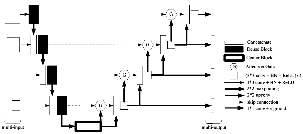
[0032] The present invention provides a thyroid ultrasound image segmentation method based on deep learning, which mainly includes five modules including data acquisition, data preprocessing, network model construction, data training and parameter adjustment, data testing and evaluation, such as figure 1 shown. The specific implementation steps are as follows:

[0033] 1. To preprocess the original ultrasound image, divide the training set and verification set;

[0034] 1) Remove patient privacy information and imaging equipment annotations on ultrasound images;

[0035] 2) Data labels are made by a professional team of ultrasound imaging physicians;

[0036] 3) Divide the original data into training set, verification set and test set according to the ratio of 6:2:2, and the labels are the same;

[0037] 4) The image resolution is unified to 256*256; and the ...


PUM


Abstract

The invention belongs to the technical field of medical image processing and computer vision, and particularly relates to a medical image segmentation method based on deep learning. According to the method, on the basis of U-Net Baseline, multiple technologies such as a multi-scale framework, a dense convolutional network, an attention mechanism, a pyramid model and small sample enhancement are fused, the method is helpful for realizing feature reuse, recovering lost context information, inhibiting response of an unrelated region and improving the performance of a small ROI, solves the problems of few ultrasonic image samples, low pixel, fuzzy boundary, large difference and other pain points, and obtains an optimal segmentation effect.

Description

technical field [0001] The invention belongs to the technical field of medical image processing and computer vision, and in particular relates to a medical image segmentation method based on deep learning. Background technique [0002] With the development of technology, doctors began to use a large number of medical image data as the basis for medical diagnosis and treatment, thus promoting the development and progress of various new technologies. However, with the development of technology, how to correctly segment medical images has become an important bottleneck restricting the development of various technologies. It can even be said that accurate image segmentation has become the most basic and important problem in the field of medical images and needs to be solved urgently. [0003] In recent years, with the continuous improvement of computing power and the continuous increase of data volume, deep learning has made remarkable progress in the field of medical imaging. ...

Claims


Application Information

Patent Timeline
Patent Type & Authority Applications(China)
IPC IPC(8): G06T7/00G06T7/11G06N3/04G06N3/08
CPCG06T7/0012G06T7/11G06N3/08G06T2207/20081G06T2207/20084G06N3/045
Inventor 陈俊江刘宇贾树开陈智方俊李治熹
Owner UNIV OF ELECTRONICS SCI & TECH OF CHINA
Why Eureka
Try Eureka