Ultrasonic image segmentation method

A technology of ultrasonic images and original ultrasonic images, which is applied in the field of medical image processing, can solve the problems of large differences in training samples, redundant computing resources and model parameters, and small perceptual areas, so as to achieve the optimal segmentation effect, realize extraction, improve performance effect

Pending Publication Date: 2020-05-15
UNIV OF ELECTRONIC SCI & TECH OF CHINA +1
View PDF3 Cites 6 Cited by
  • Summary
  • Abstract
  • Description
  • Claims
  • Application Information

AI Technical Summary

Problems solved by technology

[0004] Most of the current medical image segmentation algorithms are based on U-Net (U-Net Baseline). However, due to the problems of large differences between training samples and small perceptual regions (ROI) in medical images, there is a gap betwe

Method used


Image

  • Ultrasonic image segmentation method
  • Ultrasonic image segmentation method
  • Ultrasonic image segmentation method

Examples


Embodiment Construction

[0029] Below in conjunction with accompanying drawing and emulation the present invention is described in detail:

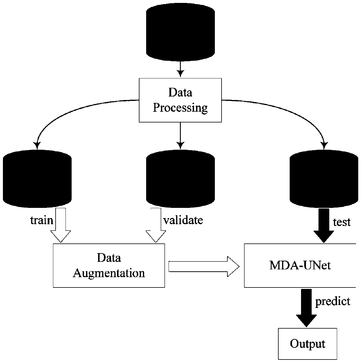
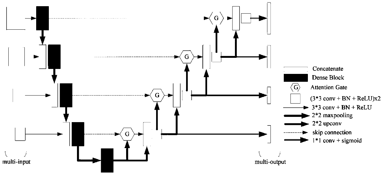
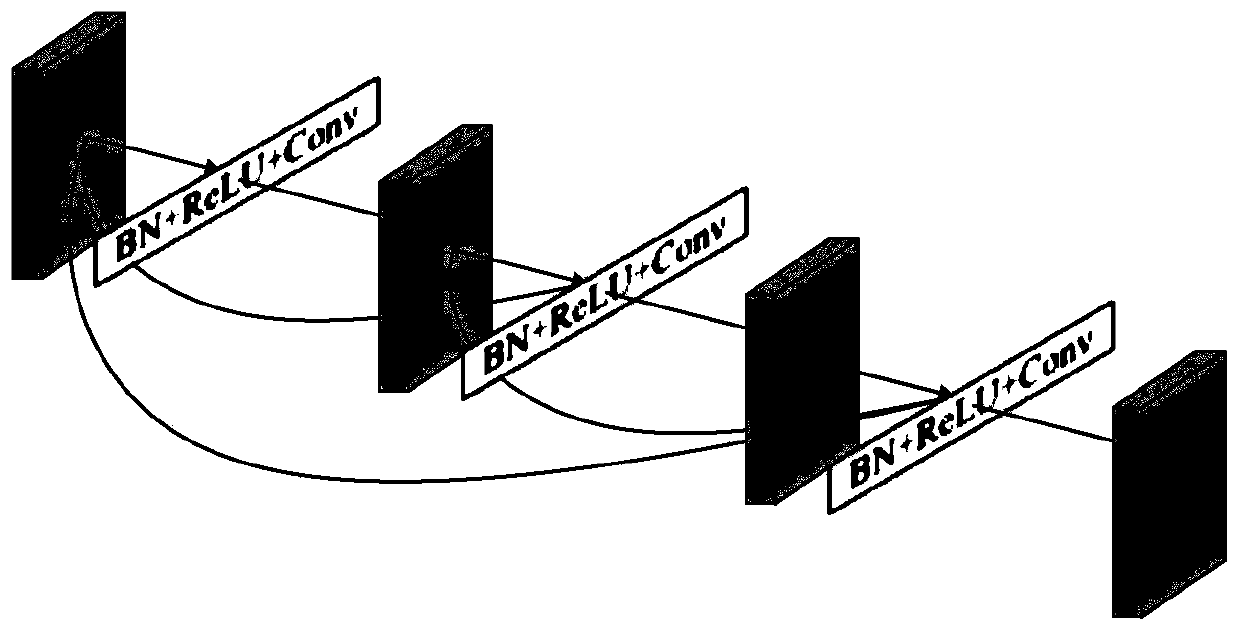
[0030] The present invention provides a thyroid ultrasound image segmentation method based on deep learning, which mainly includes five modules including data acquisition, data preprocessing, network model construction, data training and parameter adjustment, data testing and evaluation, such as figure 1 shown. The specific implementation steps are as follows:

[0031] 1. Preprocess the original ultrasound image, divide the training set, verification set, and test set

[0032] 1) Remove patient privacy information and imaging equipment annotations on ultrasound images;

[0033] 2) Data labels are made by a professional team of ultrasound imaging physicians;

[0034] 3) Divide the original data into training set, verification set and test set according to the ratio of 6:2:2, and the labels are the same;

[0035] 4) The image resolution is unified to 256*256; a...


PUM


Abstract

The invention belongs to the technical field of medical image processing, and particularly relates to an ultrasonic image segmentation method. Based on U-Net Baseline,. multiple technologies such as amulti-scale framework, a dense convolution network, an attention mechanism and small sample enhancement are fused, multi-dimensional feature extraction, irrelevant region response inhibition and small ROI performance improvement are facilitated, problems of few ultrasonic image samples, low pixels and fuzzy boundaries are solved, and the optimal segmentation effect is acquired.

Description

technical field [0001] The invention belongs to the technical field of medical image processing, and in particular relates to an ultrasonic image segmentation method. Background technique [0002] With the development of technology, doctors began to use a large number of medical image data as the basis for medical diagnosis and treatment, thus promoting the development and progress of various new technologies. How to correctly segment medical images has become an important bottleneck restricting the development of various technologies. It can be said that accurate image segmentation has become the most basic and important problem in the field of medical images and needs to be solved urgently. [0003] In recent years, with the improvement of computing performance and the increase of data volume, deep learning has made remarkable progress in the field of medical imaging. Convolutional neural network (CNN) can capture the nonlinear mapping between input and output, and automa...

Claims


Application Information

Patent Timeline
IPC IPC(8): G06T7/10
CPCG06T7/10G06T2207/10132G06T2207/20081G06T2207/20084
Inventor 陈俊江刘宇贾树开陈智方俊梁羽
Owner UNIV OF ELECTRONIC SCI & TECH OF CHINA
Why Eureka
Try Eureka