
Low-dose PET three-dimensional reconstruction method based on deep learning

A low-dose, PET scanner technology, applied in the field of medical imaging, can solve the problems of insufficient generalization ability of neural networks, performance bottlenecks, and inability to fully retain, and achieves suppression of insufficient generalization ability, reduction of required time, and high signal-to-noise. the effect of

Active Publication Date: 2020-06-23
ZHEJIANG LAB +1
View PDF16 Cites 25 Cited by
  • Summary
  • Abstract
  • Description
  • Claims
  • Application Information

AI Technical Summary

Problems solved by technology

Because the traditional low-dose PET reconstruction process cannot fully preserve the effective high-frequency information in the original data, the generalization ability of the neural network used to fit the mapping is insufficient, resulting in image artifacts and quantitative errors, which have become the performance bottleneck of this type of technical route

Method used


Image

  • Low-dose PET three-dimensional reconstruction method based on deep learning
  • Low-dose PET three-dimensional reconstruction method based on deep learning
  • Low-dose PET three-dimensional reconstruction method based on deep learning

Examples


Embodiment Construction

[0020] The content of the present invention will be further elaborated below in conjunction with the accompanying drawings and embodiments.

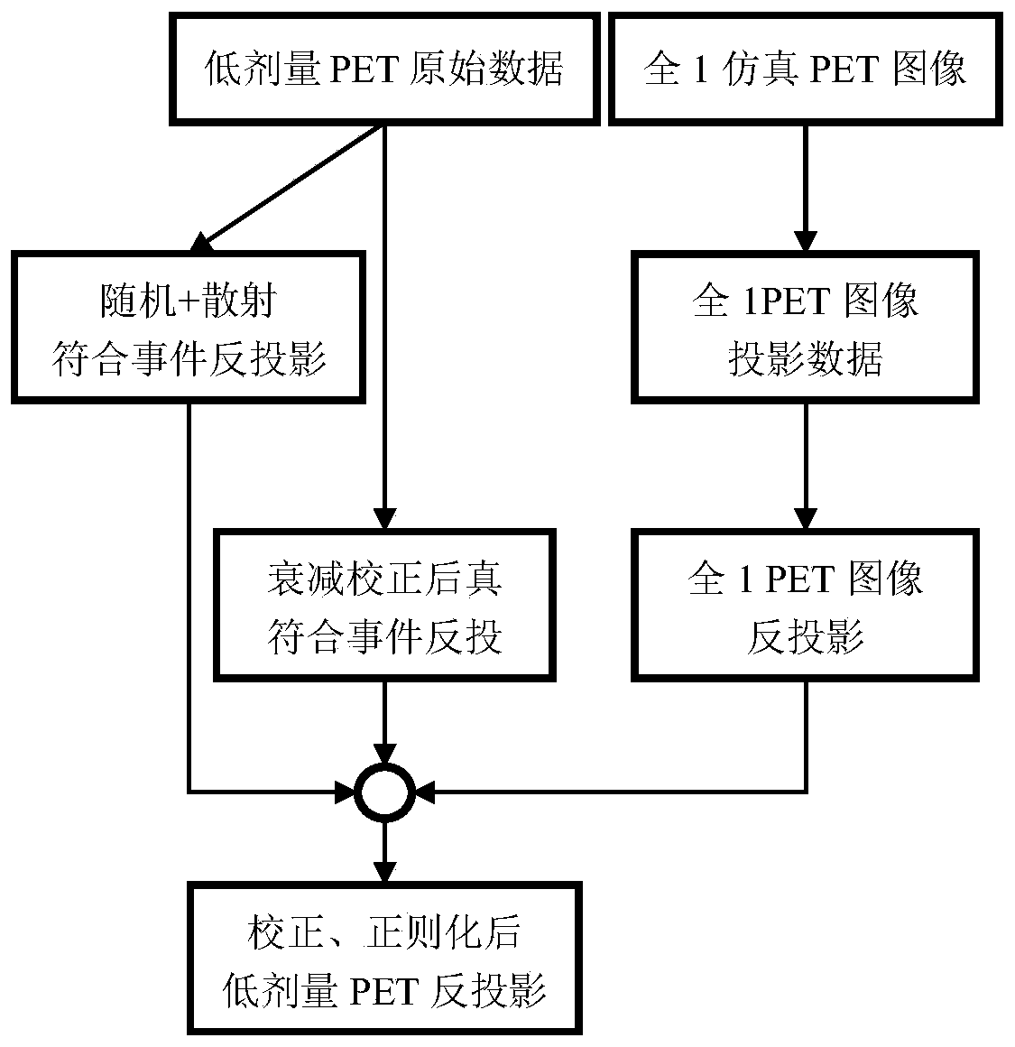
[0021] The low-dose raw data received by the PET scanner is a three-dimensional sinogram formed by the projection of the PET image through three-dimensional X-ray transformation. Since the three-dimensional sinogram not only includes the axial plane projection, but also includes the oblique plane projection passing through the axial plane, it has the characteristics of large amount of data and highly redundant information, and the direct use of neural network to fit the mapping between sinogram and PET image is limited. Due to the limitations of computer computing and storage capabilities, it is difficult to realize. The present invention proposes a lossless back-projection method for a three-dimensional sinogram, the flow chart of which is as follows figure 1 Shown:

[0022] (1.1) The low-dose PET raw data is processed by attenuation ...


PUM


Abstract

The invention discloses a low-dose PET three-dimensional reconstruction method based on deep learning. The method comprises the following steps: carrying out lossless back projection on low-dose PET original data to an image domain; an appropriate three-dimensional deep neural network structure is selected to fit mapping between low-dose PET back projection and standard-dose PET images; after training sample learning and network parameter fixing, PET image three-dimensional reconstruction starting from low-dose PET original data is realized, so that a low-dose PET reconstructed image which islower in noise and higher in resolution compared with a traditional reconstruction algorithm and image domain noise reduction processing is obtained.

Description

technical field [0001] The invention relates to the field of medical imaging, in particular to a method for three-dimensional reconstruction of low-dose PET based on deep learning. Background technique [0002] Positron Emission Tomography (PET) is a medical image that can provide biochemical and quantitative physiological information in vivo. It has important applications in oncology, cardiology, neurology, and mental diseases. PET / CT is also It has become the internationally recognized gold standard for tumor detection. The PET imaging process includes injecting a radioactive tracer into the patient before scanning. The tracer decays to generate positrons when it participates in physiological metabolism. The annihilation effect of the positrons and adjacent electrons produces 511keV photon pairs that move inversely. The photon hits the receiver of the PET scanner to form a certain number of lines of response (Line of Response, LOR), and saves it as a three-dimensional PET...

Claims


Application Information

Patent Timeline
Patent Type & Authority Applications(China)
IPC IPC(8): G06T5/00G06T11/00G06T17/00G06N3/08G06N3/04
CPCG06T17/00G06T11/005G06N3/08G06T2207/10104G06N3/045G06T5/70G06T11/006G06T2211/441G06T2211/421
Inventor 朱闻韬杨宝周龙叶宏伟陈凌饶璠王瑶法
Owner ZHEJIANG LAB
Learn More