Eureka AIR delivers breakthrough ideas for toughest innovation challenges, trusted by R&D personnel around the world.

Multi-modal endoscope and endoscopic imaging system

An imaging system and multi-modal technology, applied in the field of medical devices, can solve the problem of inability to provide tomographic images of tissues and organs, and achieve the effect of poor imaging resolution

Inactive Publication Date: 2020-07-17
SHANGHAI INST OF TECH
View PDF7 Cites 3 Cited by
  • Summary
  • Abstract
  • Description
  • Claims
  • Application Information

AI Technical Summary

Problems solved by technology

This design can perform real-time imaging, but it cannot perform multi-modal imaging, so it cannot provide more accurate tomographic images of tissues and organs

Method used


Image

  • Multi-modal endoscope and endoscopic imaging system
  • Multi-modal endoscope and endoscopic imaging system
  • Multi-modal endoscope and endoscopic imaging system

Examples


Embodiment

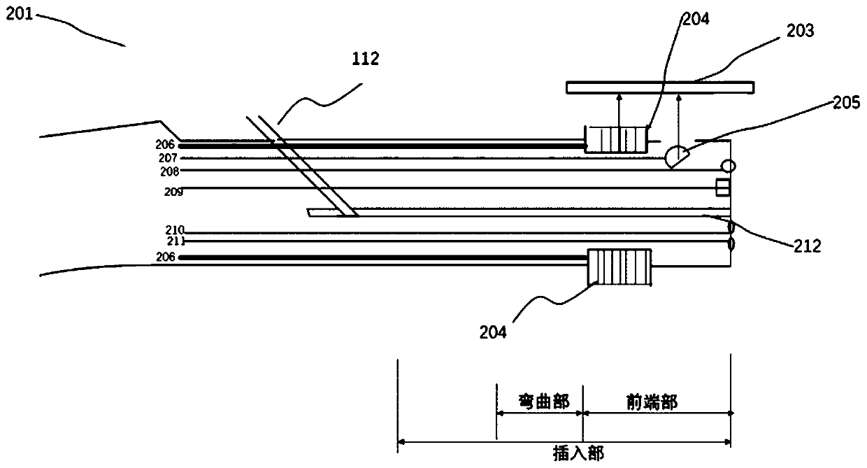
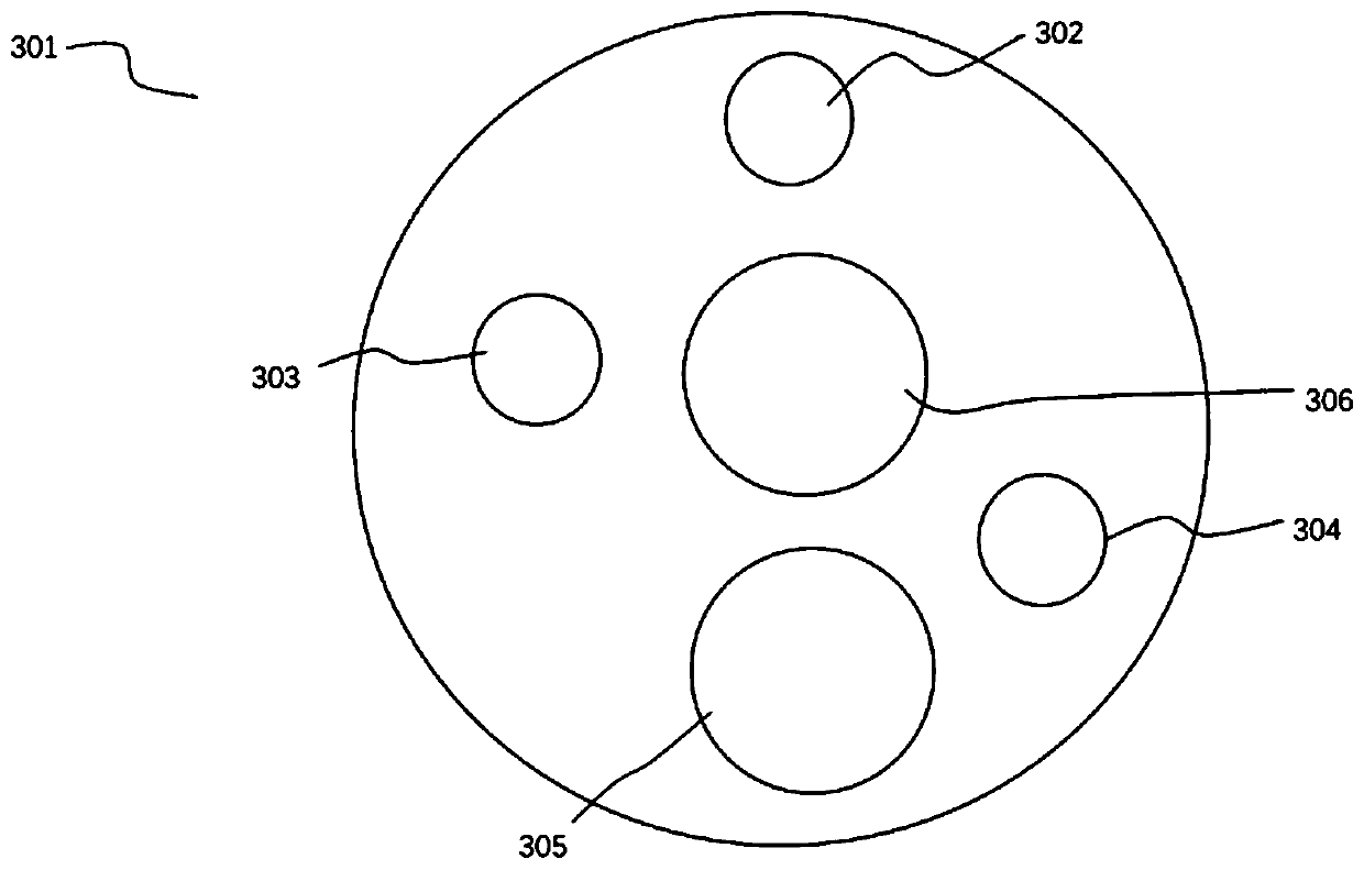
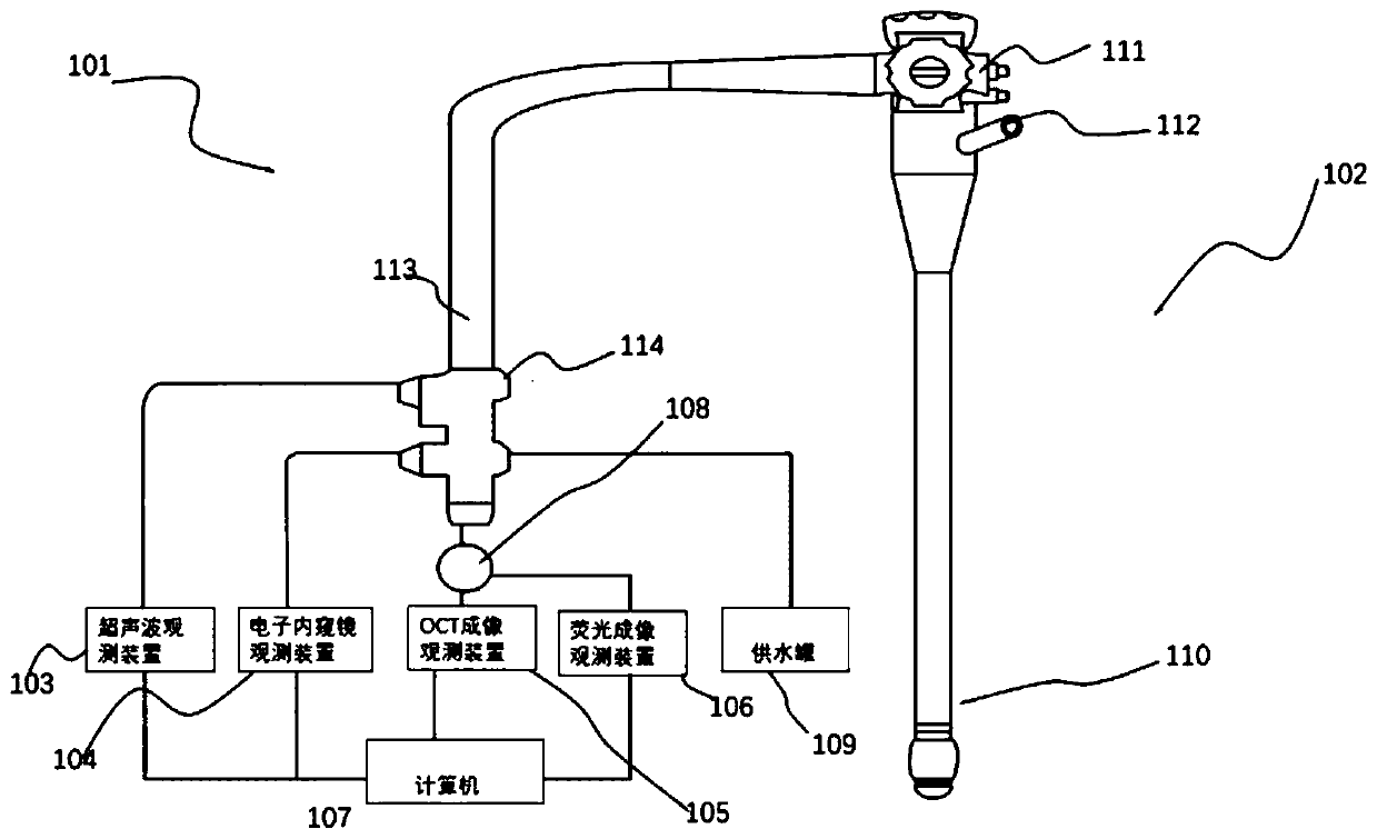
[0042] The invention relates to a multi-mode endoscope. The lens of the multi-mode endoscope can convert acoustic signals-electrical signals, optical signals-electrical signals through signal lines and optical fibers. It has comprehensive in-body, tomographic imaging, Clinical features in several aspects of high resolution and imaging depth, this endoscope includes:

[0043] The insertion part, the insertion part, has a front end, which is a top hard structure, and the front end includes a plurality of ultrasonic transducers, probes, and imaging elements such as CCD / CMOS for receiving optical signals arranged in lateral rows. The imaging device is inserted into the digestive tract (esophagus, stomach, duodenum, large intestine) or respiratory tract (trachea, bronchi) of the subject, and can image the digestive tract and respiratory tract. At the same time, the insertion part also has a bending part and a flexible tube part. The bending part is located at the rear side of the f...


PUM


Abstract

The invention relates to a multi-modal endoscope and an endoscopic imaging system. The system combines ultrasonic imaging, optical coherence tomography, fluorescence imaging and electronic endoscopicimaging are combined, has a photographing optical system and photographing elements, the photographing elements are inserted into digestive tracts (esophagus, stomach, duodenum and large intestine) orrespiratory tracts (trachea and bronchus) of an object, and can photograph the digestive tracts and respiratory tracts; and an optical coherence imaging device and a fluorescence imaging device withdifferent imaging principles are arranged, the two devices are in signal connection with an image processing device, and a wavelength division multiplexer is combined and used as a fluorescence imaging system, so that the advantages of various imaging devices can be combined to obtain medical images of tissue and organs, and the system helps to capture more accurate tomograms of the tissue and organs. Compared with the prior art, the multi-modal endoscope and endoscopic imaging system provided by the invention have excellent performance.

Description

technical field [0001] The invention relates to the technical field of medical instruments, in particular to a multi-mode endoscope and an endoscope imaging system. Background technique [0002] In recent years, the continuous development of new imaging technologies has provided new means for early diagnosis of diseases, including endoscopic ultrasound imaging, optical coherence tomography, fluorescence imaging, confocal imaging and so on. [0003] Endoscopic Ultrasonography System (EUS) is a medical device that integrates ultrasound and endoscope. After the curved portion of the ultrasonic endoscope is inserted into the human body, the endoscopic probe at the front end performs tomographic scanning of the internal organs, thereby obtaining ultrasonic images of the internal organs. Due to the deep detection depth of ultrasonic imaging, ultrasonic endoscopy plays a very important role in the diagnosis and treatment of endoscopy rooms in large domestic hospitals. [0004] Op...

Claims


Application Information

Patent Timeline
Patent Type & Authority Applications(China)
IPC IPC(8): A61B1/005A61B1/012A61B1/04A61B5/00A61B8/08
CPCA61B1/005A61B1/04A61B1/00131A61B1/012A61B5/0066A61B5/0071A61B8/0833
Inventor 戴翠霞杜奉献孔瑞明邱锐高磊葛坚坚董柏文
Owner SHANGHAI INST OF TECH
Why Eureka
Eureka Blog
Learn More