Retinal Vascular Image Segmentation System Based on Global Information Convolutional Neural Network

A convolutional neural network and retinal blood vessel technology, applied in the field of medical image processing, can solve the problems of limited use of global information and easy loss of important features, and achieve good universality and good performance.

Active Publication Date: 2021-02-09
HARBIN INST OF TECH
View PDF7 Cites 0 Cited by
  • Summary
  • Abstract
  • Description
  • Claims
  • Application Information

AI Technical Summary

Problems solved by technology

[0006] The purpose of the present invention is to solve the problem of limited utilization of global information and easy loss of important features in the existing convolutional neural network retinal vessel image segmentation, and now proposes a retinal vessel image segmentation system based on global information convolutional neural network

Method used


Image

  • Retinal Vascular Image Segmentation System Based on Global Information Convolutional Neural Network
  • Retinal Vascular Image Segmentation System Based on Global Information Convolutional Neural Network
  • Retinal Vascular Image Segmentation System Based on Global Information Convolutional Neural Network

Examples


specific Embodiment approach 1

[0024] Specific Embodiment 1: In this embodiment, the retinal vessel image segmentation system based on the global information convolutional neural network includes:

specific Embodiment approach

[0026] The DRIVE public dataset contains 40 color retinal images, 7 of which are lesion images; all fundus images in DRIVE have a resolution of 565×584 pixels; the dataset is evenly divided into a training set and a test set;

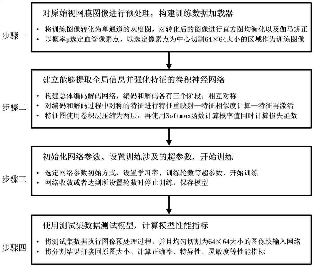
[0027] Preprocess the original retinal image and build a training data loader;

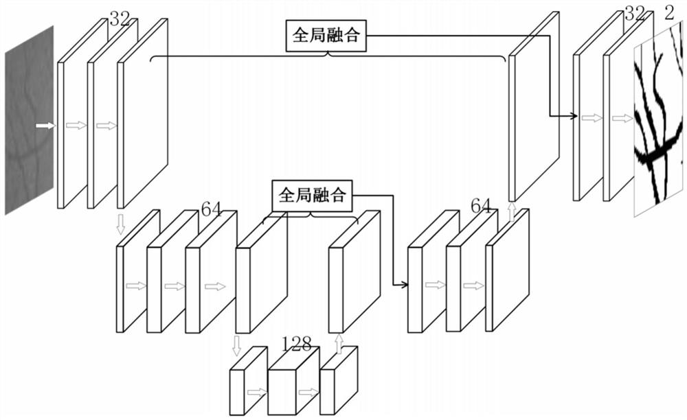
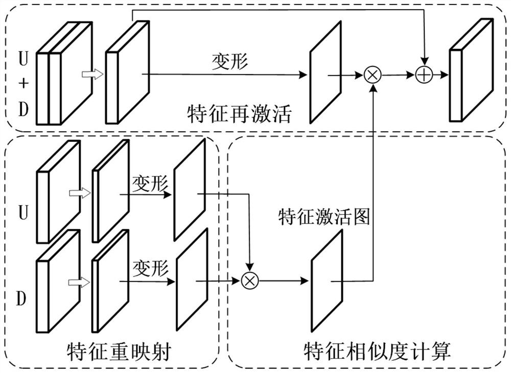
[0028] Establish a convolutional neural network that can extract global information and strengthen features. The overall network structure uses the framework of encoding first and then decoding. During the decoding process, the feature map is remapped → feature similarity calculation → feature reactivation to complete the global Integration of information and enhancement of vulnerable features;

[0029] Initialize network parameters, set hyperparameters involved in training neural network locks, and start training;

[0030] Use the trained model to test and calculate performance metrics;

[0031] The flow chart of the present invention is as figure 1 As shown, the detail...

specific Embodiment approach 2

[0037] Specific embodiment 2: The difference between this embodiment and specific embodiment 1 is that the image processing main module is used to collect the original retinal image, preprocess the collected original retinal image, obtain the processed image, and preprocess the The final image is input to the training main module and the detection main module; the specific process is:

[0038] The DRIVE public dataset contains 40 color retinal images, 7 of which are lesion images; all fundus images in DRIVE have a resolution of 565×584 pixels; the dataset is evenly divided into a training set and a test set;

[0039] A1. Obtain training data and read 20 images of the training set. These 20 images are RGB three-channel color images, and these 20 training images are converted into single-channel grayscale images. For the converted image retinal background and For the problem that the contrast between blood vessels is not obvious enough, perform histogram equalization on the conv...


PUM


Abstract

Retinal Vascular Image Segmentation System Based on Global Information Convolutional Neural Network. The invention relates to a retinal vessel image segmentation system. The invention aims to solve the problems of limited utilization of global information and easy loss of important features in the existing convolutional neural network retinal vessel image segmentation. The system of the present invention includes: an image processing main module, a neural network main module, a training main module and a detection main module; the image processing main module is used to collect original retinal images, preprocess the collected original retinal images, and process The final image is input into the training main module and the detection main module; the neural network main module is used to establish a convolutional neural network capable of extracting global information and strengthening features; the training main module is used to initialize network parameters and obtain a trained volume A product neural network model; the detection main module is used to use the trained model to test and calculate the model performance index. The invention belongs to the field of retinal blood vessel image segmentation system.

Description

technical field [0001] The invention belongs to the field of medical image processing, in particular to a retinal blood vessel image segmentation system. Background technique [0002] Fundus images contain rich pathological information. By observing the length, width, and curvature of retinal blood vessels, doctors can diagnose various cardiovascular and ophthalmic diseases, such as glaucoma, hypertension, and arteriosclerosis. Because fundus images can be easily obtained by fundus cameras without complex procedures such as angiography, they are widely used in clinical disease analysis. In order to more accurately and quantitatively assess related diseases, proper retinal vessel image segmentation is required to eliminate interference from other tissues of the eye. High-precision automatic retinal vessel image segmentation technology has broad application prospects. Existing retinal image segmentation techniques are mainly divided into the following three categories. One ...

Claims


Application Information

Patent Timeline
Patent Type & Authority Patents(China)
IPC IPC(8): G06T7/11G06N3/04G06N3/08
CPCG06T7/11G06N3/08G06T2207/30041G06T2207/30101G06N3/045
Inventor 李翔罗浩李明磊蒋宇辰李款尹珅
Owner HARBIN INST OF TECH
Why Eureka
Try Eureka