Unlock instant, AI-driven research and patent intelligence for your innovation.
Bioanalysis chip capable of realizing cell capture and fixation
What is Al technical title?
Al technical title is built by PatSnap Al team. It summarizes the technical point description of the patent document.
A bio-analysis and bio-molecular technology, applied in analytical materials, biological testing, material excitation analysis, etc., can solve problems such as cell loss, and achieve the effect of less loss
Pending Publication Date: 2020-10-16
DALIAN INST OF CHEM PHYSICS CHINESE ACAD OF SCI
View PDF4 Cites 4 Cited by
Summary
Abstract
Description
Claims
Application Information
AI Technical Summary
This helps you quickly interpret patents by identifying the three key elements:
Problems solved by technology
Method used
Benefits of technology
Problems solved by technology
Due to the limitations of conventional single-cell analysis, during the process of single-cell protein analysis, the cell loss is serious, and it is difficult to perform multiple secretion detection operations on the same cell continuously
Method used
Image
Click on the blue label to locate the original text in one second.
Reading with bidirectional positioning of images and text.
Examples
Embodiment 1
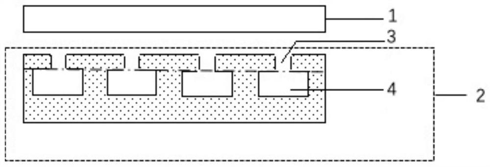
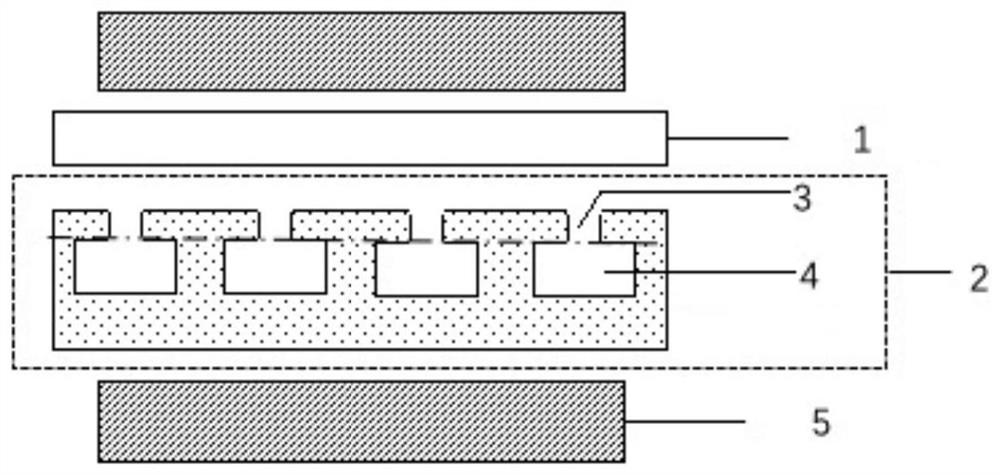
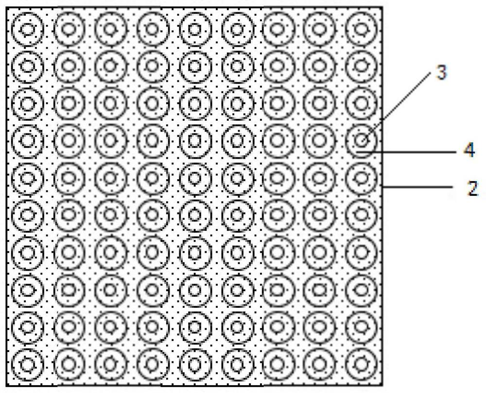
[0051] A chip for biological analysis of cell capture and immobilization, such as Figure 1 ~ Figure 3 shown. The chip includes a two-layer structure, which is a biological analysis substrate 1, a microwell array chip 2, and the micropore opening 3 and the micropore cavity 4 of the microwell array chip 2 are respectively manufactured, and combined by thermal bonding to obtain a complete microwell array chip 2 .
[0052] The microwell array chip 2 is a PDMS microwell array chip.
[0053] In this embodiment, the PDMS microwell array chip is completed using MEMS technology and replication molding technology, such as Figure 4 , Figure 5 As shown, it specifically includes the following steps:
[0054] Step 1: Mix PDMS (Gel A and Gel B (PDMS cross-linking agent) at a mass ratio of 10:1, stir evenly, and place in a vacuum desiccator to degas until there are no bubbles;
[0055] Step 2: Pour PDMS onto the micropore opening 3 silicon template 6, and use a glue homogenizer to sha...
Embodiment 2
[0060] The chip in this example uses U937 (human histiocytic lymphoma cells, ATCC-CRL-1593.2) suspension cells and scc6 (oral squamous cell carcinoma) adherent cells to verify the microwell array chip of Example 1 at multiple time points When testing, the cell retention status. The experimental process of this embodiment is as follows Figure 6 shown, including the following steps:
[0061] Step 1: Prepare multiple clean slides and stain the cells with calcein;
[0062] Step 2: Treat the PDMS microwell array chip with PLASMA for hydrophilic treatment;
[0063] Step 3: Divide the above two types of cells into 1×10 5 cells / ml, 400μl / chip were inoculated on PDMS microwell array chips;
[0064] Step 4: Cover with a clean glass slide and fix the chip with a clamp;
[0066] Step 6: Remove the fixture, place the chip in the incubator for 2 hours, cover it with a clean glass slide, and fix the chip with the fixtu...
Embodiment 3
[0070] A chip for biological analysis of cell capture and immobilization, such as Figure 1 ~ Figure 3 shown. The chip includes a two-layer structure, followed by a biological analysis substrate 1, and a narrow-mouth and wide-bottom microwell array chip. Wide bottom microwell array chip4.
[0071] The biological analysis chip 4 is a PDMS microwell array chip.
PUM
Property
Measurement
Unit
height
aaaaa
aaaaa
height
aaaaa
aaaaa
height
aaaaa
aaaaa
Abstract
The invention relates to the fields of micro-fluidic chips, cell operation and biological analysis, in particular to a bioanalysischip capable of realizing cell capture and fixation. The bioanalysischip comprises a bioanalysis substrate and a micropore array chip, the micropore array chip comprises a substrate and a micropore array formed by a plurality of micropores on the substrate, and the inner diameter of micropore openings of the micropores is smaller than that of micropore cavities. According to the micropore structure, the diameter of the opening of the micropore is slightly larger than the diameter of cells, single cell capture can be achieved, the cell is not prone to loss after entering the micropore cavity, the bioanalysis chip can be used for multi-time-point detection of the same cell, and the micropore cavity can provide sufficient space and culture solution for long-time culture of various cells. The bioanalysis chip can be used for cell multi-index biomolecular analysis, cell multi-index multi-time-point dynamic biomolecular analysis and single-cell multi-index multi-time-point biomolecular analysis, such as secretory proteinspectrum analysis and exosome analysis.
Description
technical field [0001] The invention relates to the fields of microfluidic chip, cell operation and biological analysis, in particular to a biological analysis chip capable of capturing and fixing cells. Background technique [0002] The most challenging part of single-cell analysis is single-cell capture. The single-cell capture procedure on the microfluidic chip should be simple and efficient, and can maintain single-cell activity. At present, the existing single cell capture technology can be roughly divided into two types: one is not applied by external force, and the cells in the cell suspension are captured by the microstructure on the chip, such as hydrodynamic method and microfluidic method; External forces such as light, sound, electricity, magnetism, etc. drive cell movement for capture, such as dielectrophoresis. With the existing single-cell capture technology, it is difficult to perform long-term culture and dynamic detection on the premise of ensuring the acti...
Claims
Application Information
Patent Timeline
Application Date:The date an application was filed.
Publication Date:The date a patent or application was officially published.
First Publication Date:The earliest publication date of a patent with the same application number.
Issue Date:Publication date of the patent grant document.
PCT Entry Date:The Entry date of PCT National Phase.
Estimated Expiry Date:The statutory expiry date of a patent right according to the Patent Law, and it is the longest term of protection that the patent right can achieve without the termination of the patent right due to other reasons(Term extension factor has been taken into account ).
Invalid Date:Actual expiry date is based on effective date or publication date of legal transaction data of invalid patent.
