Dual fluorescence imaging method of mucoprotein 1 and sialyl glycosyl thereof and application

A technology of sialic acid and mucin, which is applied in the field of fluorescence imaging and biomedicine, can solve the problems of complex analysis and lack of specificity, and achieve high specificity and biocompatibility, low cytotoxicity, and strong fluorescence penetration ability Effect

Active Publication Date: 2020-11-27
JIANGNAN UNIV
View PDF7 Cites 1 Cited by
  • Summary
  • Abstract
  • Description
  • Claims
  • Application Information

AI Technical Summary

Problems solved by technology

Clinical diagnosis is usually made by combining various biochemical and imaging techniques, such as enzyme-linked immunosorbent assay (ELISA), hematoxyli

Method used


Image

  • Dual fluorescence imaging method of mucoprotein 1 and sialyl glycosyl thereof and application
  • Dual fluorescence imaging method of mucoprotein 1 and sialyl glycosyl thereof and application
  • Dual fluorescence imaging method of mucoprotein 1 and sialyl glycosyl thereof and application

Examples


Embodiment 1

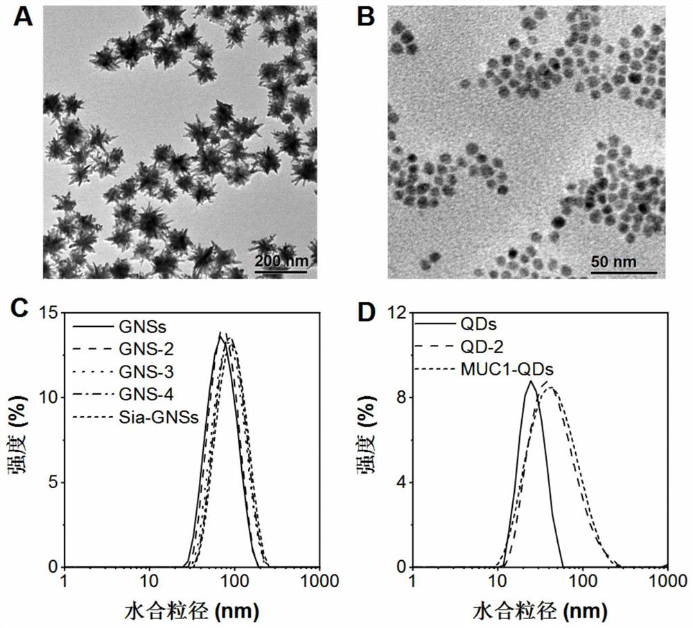
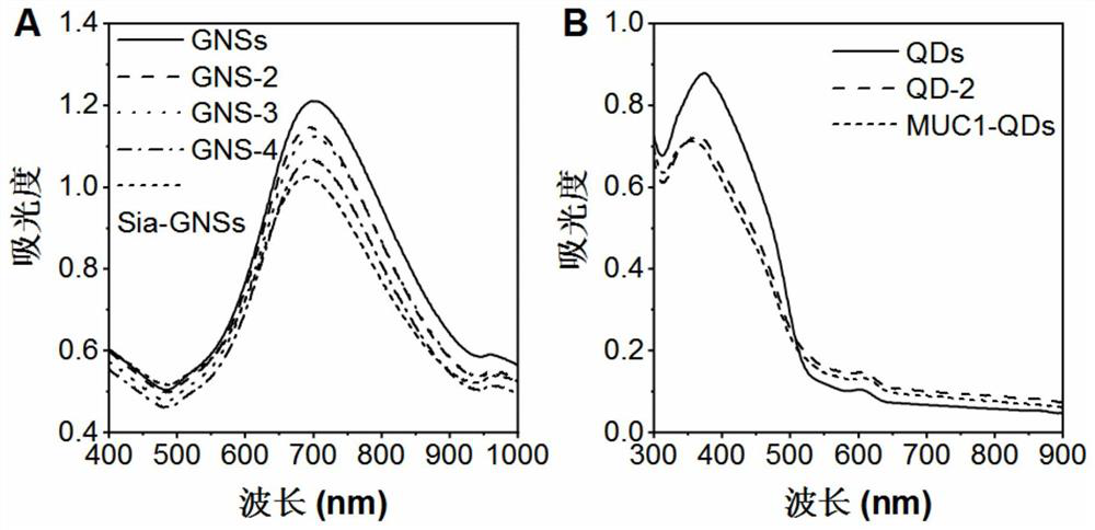
[0048] Embodiment 1: the synthesis of Sia-GNSs nanoprobe

[0049] (1) Synthesis of gold nanostars GNSs (gold nanostars)

[0050] All glass instruments were soaked in aqua regia, washed with double distilled water, and dried for later use. Gold nanostars were prepared by seed crystal growth method. The water used in the following steps is 18.2 MΩ Milli-Q ultrapure water.

[0051] The synthesis steps of gold nanostars are:

[0052] (1) Synthesis of seed crystals: 100mL of 1mM tetrachloroauric acid trihydrate is placed in a reaction flask, heated and stirred at 100°C until boiling, then 15mL of sodium citrate solution with a concentration of 10g / L is added, and boiled for 15 minutes to make The color of the solution changed from light yellow to wine red. Cool to room temperature, filter with a 0.22 μm membrane filter, and store in a refrigerator at 4°C.

[0053] (2) Preparation of gold nanostars: After the seed crystal solution in step (1) is synthesized, the gold nanostars ...

Embodiment 2

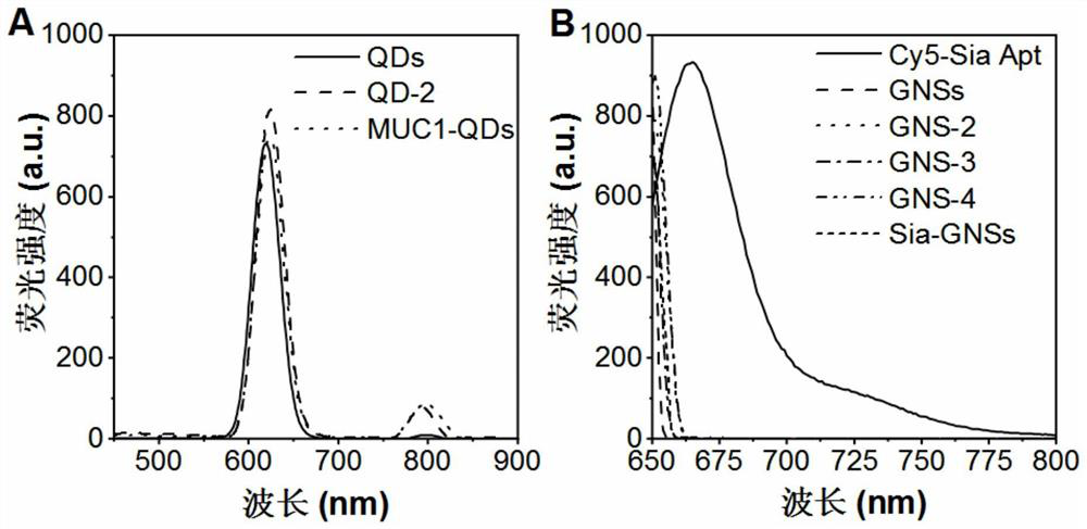
[0061] Embodiment 2: the synthesis of MUC1-QDs nanoprobe

[0062] (1) Quantum dots and MUC1 aptamers are coupled to synthesize QD-2:

[0063] Carboxyl water-soluble CdSe@ZnS quantum dots were purchased from Wuhan Jiayuan Quantum Dot Technology Development Co., Ltd. Add 4 μL of EDC with a concentration of 0 mg / mL and 1.6 μL of NHS with a concentration of 10 mg / mL to 80 μL of PBS solution containing 2 μM QDs, react at room temperature for 1 hour, and then add 80 μL of MUC1 aptamer with a concentration of 100 μM (sequence: 5'-NH 2 -C 6 -GCAGTTGATCCTTTGGATACCCTGG-3'), mix well, shake at room temperature for 2 hours. Centrifuge at 13,000 rpm for 10 minutes, and then disperse in 80 μL of PBS to obtain QD-2.

[0064] (2) QD-2 and the aminated polyethylene glycol (NH 2 -PEG2000) coupling to synthesize MUC1-QDs:

[0065] Add 4 μL of 10 mg / mL EDC and 1.6 μL of 10 mg / mL NHS to the QD-2 prepared in step (1), react at room temperature for 1 hour, and then add 10 μL of 10 mg / mL NHS 2...

Embodiment 3

[0067] Embodiment 3: the stability test of nanoprobe

[0068] Stability is one of the most important properties of nanocarriers. Nanoparticles used in the field of biomedicine must be able to be stably dispersed in a certain concentration range of salt solution or medium. Dilute the nanoprobe Sia-GNSs prepared in Example 1 and the nanoprobe MUCl-QDs prepared in Example 2 into aqueous solution, phosphate buffer (PBS, pH=7.4) and high-glucose medium containing 10% fetal bovine serum (DMEM), the concentration after dilution is 0.1nM, and its particle size change within 7 days is measured by DLS, so as to judge its stability. The result is as Figure 4 As shown, there is no obvious particle size change within 7 days, indicating that the two nanoprobes have good stability.


PUM

PropertyMeasurementUnit
Concentrationaaaaaaaaaa
Particle sizeaaaaaaaaaa
Thicknessaaaaaaaaaa

Abstract

The invention discloses a dual fluorescence imaging method of mucoprotein 1 and sialyl glycosyl thereof and application, and belongs to the technical field of fluorescence imaging and the field of biomedicine. According to the invention, a sialic acid-gold nanostar probe Sia-GNSs and a mucoprotein 1-quantum dot probe MUC1-QDs, which are high in specificity and biocompatibility, are successfully constructed. MUC1 in the MCF-7 cell can be specifically recognized by using the Sia-GNSs and the MUC1-QDs in a combined manner. The nano probe prepared by the invention can realize dual fluorescence imaging and relative quantification of MUC1 protein skeleton and Sia glycosyl thereof in vitro, in vivo and in clinical breast cancer patient tissue samples. The invention provides a simple and effectiveplatform, and has huge potential in clinical cancer diagnosis.

Description

technical field [0001] The invention relates to a double fluorescence imaging method and application of mucin 1 and its sialyl glycosyl, belonging to the technical field of fluorescence imaging and the field of biomedicine. Background technique [0002] Cancer is a major cause of disease burden, and cancer detection and imaging for clinical use is extremely important for early cancer diagnosis, monitoring recurrence, and drug discovery and development. Clinical diagnosis is usually made by combining various biochemical and imaging techniques, such as enzyme-linked immunosorbent assay (ELISA), hematoxylin-eosin (H&E) staining, computed tomography (CT), or magnetic resonance imaging (MRI), This complicates the analysis and leads to a lack of specificity. The detection of protein biomarkers can be used for the identification and monitoring of cancer. Mucin 1 (MUC1) is a highly glycosylated type I transmembrane O-glycoprotein that is abnormally expressed on the surface of a va...

Claims


Application Information

Patent Timeline
IPC IPC(8): G01N33/68G01N33/574G01N33/58G01N33/53G01N21/64
CPCG01N21/6402G01N21/6428G01N21/6458G01N33/5308G01N33/574G01N33/57492G01N33/582G01N33/587G01N33/588G01N33/68G01N2333/4725
Inventor 尹健王晓丽胡静叶雨霏
Owner JIANGNAN UNIV
Why Eureka
Try Eureka