Mouse bone marrow dendritic cell induction and purification method

A dendritic cell and purification method technology, applied in the field of mouse bone marrow dendritic cell induction and purification, can solve the problems of small number, complicated operation, long dendritic cell induction period, etc., and achieve the effect of cost reduction

Active Publication Date: 2020-12-01
SOUTHEAST UNIV
View PDF5 Cites 1 Cited by
  • Summary
  • Abstract
  • Description
  • Claims
  • Application Information

AI Technical Summary

Problems solved by technology

[0003] At present, in the process of studying DC cells, there are various induction and purification methods of mouse bone marrow-derived dendritic cells, but there are shortcomings such as long induction period of dendritic cells, small number, and high purification cost, which make it difficult to meet the basic requirements. For the needs of research or clinical research, in order to change the above shortcomings, the induction time is extended to increas...

Method used


Image

  • Mouse bone marrow dendritic cell induction and purification method
  • Mouse bone marrow dendritic cell induction and purification method
  • Mouse bone marrow dendritic cell induction and purification method

Examples


Embodiment 1

[0028] A method for inducing and purifying mouse bone marrow dendritic cells, comprising the following steps:

[0029] Extraction of S1 mouse bone marrow cells:

[0030] S101. Select 4-week-old mice to be killed, soak them in 75% alcohol and put them into an ultra-clean table. After 3-5 minutes, take them out and put them into a 10cm-diameter sterile petri dish;

[0031] S102. Carefully pinch the abdominal skin of the iliac fossa of the mouse with sterile tweezers, carefully cut the skin with ophthalmic scissors, remove the skin from the thigh to the ankle joint from top to bottom, cut at the ankle and hip joint, and free the mouse two lower limbs;

[0032] S103. Peel off the muscles of the knee joint, and split them at the junction of the femur and tibia against the direction of joint bending, and the tibial plateau can be seen; remove the appendages of the distal end of the femur, use gauze to remove the muscles attached to the bone, and put it in a 75% alcohol-filled conta...

Embodiment 2

[0051] In step S104, the epiphysis is not reserved, and other steps are the same as in embodiment 1.

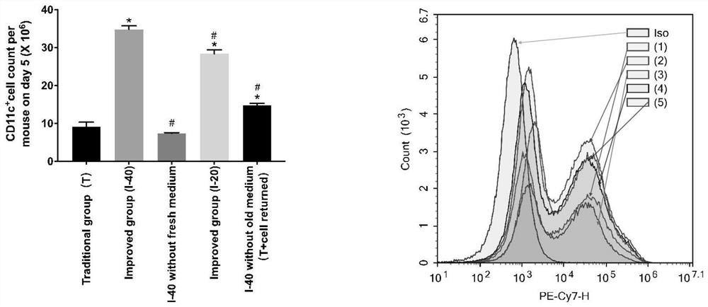
[0052] like figure 1 As shown, incubate with FVS-780 dead and alive dye for 15 minutes, and perform flow cytometry after elution with PBS. The ratio of living cells R1 accounts for more than 90% of the total cells in the P2 gate; count the number of cells in the R1 gate and calculate the total number of cells, and keep the epiphysis The number of cells in the non-retained group increased by 18%.

Embodiment 3

[0054] Traditional method group [Traditional group (T)]:

[0055] ① On day 0, according to the number of viable bone marrow cells, adjust the cell concentration to 4×10⁵ cells / mL with 10% RPMI-1640 medium containing GM-CSF, IL-4 (the final concentration is 40 ng / mL), Take 10mL of cell suspension and transfer it into a 10cm diameter petri dish (three replicate wells), and place it at 37°C with 5% CO 2 Cultivated in an incubator;

[0056] ② On the third day, completely remove the old culture medium containing cells in the dish, and add 10 mL of the above fresh culture medium containing the same inducer in ①;

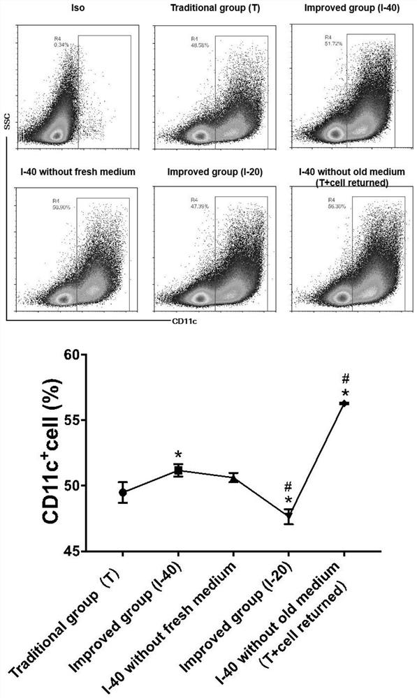
[0057] ③ On day 5, take the cell suspension in the culture dish, centrifuge at 300G for 10 minutes, wash with PBS three times, block with FCR at 4°C for 15 minutes, add CD11c flow cytometry antibody and incubate at 4°C for 30 minutes, wash with PBS three times, and measure the number of cells by flow cytometry and purity, the total amount of dendritic cells was calculate...


PUM


Abstract

The invention discloses a mouse bone marrow dendritic cell induction and purification method which comprises the following steps: S1, mouse bone marrow cell extraction: clamping and cracking reservedends of thighbone and tibia by using surgical forceps, and continuously performing washing to obtain cells to the maximum extent; S2, dendritic cell culture: keeping an original culture medium and cells on a third day of culture, and adding a fresh culture medium for continuous culture; S3, purification of dendritic cell cells: adopting Percoll stratified liquid for purification and induction. According to the invention, the epiphysis is clipped and broken by surgical forceps instead of being cut off, traditional cells discarded after centrifugation on the third day are added into a culture dish again, and an original culture medium is reserved on the basis of adding the fresh culture medium during liquid change on the third day, so that the DC number of harvested cells is greatly increased; according to the method, the purity of the obtained DC cells reaches 90% or above through Percoll liquid purification and screening, the yield is increased by 21% compared with a classic magnetic bead sorting mode, and the Percoll density sorting method is lower in cost consumption.

Description

technical field [0001] The invention relates to the field of dendritic cell culture, in particular to a method for inducing and purifying mouse bone marrow dendritic cells. Background technique [0002] Dendritic cells, also known as DC cells, are the most functional antigen-presenting cells, named for their many dendritic or pseudopodia-like protrusions when mature. DCs can synthesize a large number of MHC class II molecules; have special membrane receptors for expressing, taking up and transporting antigens; can effectively take up and process antigens and then migrate to the T cell area, with a maturation process; can activate naive T cells; a small amount Antigen and a small amount of DC are enough to activate T cells. Because of its extremely important role in the body's anti-infection immunity, anti-tumor immune response and transplant immunity, it has attracted much attention and has become a hot spot in immunology research in recent years. DCs are widely distribute...

Claims


Application Information

Patent Timeline
IPC IPC(8): C12N5/0784
CPCC12N5/0639C12N2509/00C12N2509/10C12N2501/58C12N2501/2304C12N2501/22
Inventor 鹿中华刘玲范赡文程振兴郭凤梅
Owner SOUTHEAST UNIV
Why Eureka
Try Eureka