Inhibitor for autophagy and apoptosis of retinal pigment cells RPE and application of inhibitor

A retinal pigment and inhibitor technology, applied in the field of autophagy and apoptosis inhibitors

Inactive Publication Date: 2020-12-08
TONGJI UNIV
View PDF5 Cites 3 Cited by
  • Summary
  • Abstract
  • Description
  • Claims
  • Application Information

AI Technical Summary

Problems solved by technology

However, since the current treatment methods are mostly aimed at the late stage of the disease, and all have certain side effects and prognostic risks, research on the molecular mechanism and treatment methods for the early stage of the disease is imminent

Method used


Image

  • Inhibitor for autophagy and apoptosis of retinal pigment cells RPE and application of inhibitor
  • Inhibitor for autophagy and apoptosis of retinal pigment cells RPE and application of inhibitor
  • Inhibitor for autophagy and apoptosis of retinal pigment cells RPE and application of inhibitor

Examples


Embodiment 1

[0030] Vitreous Drug Injection in SD Rats

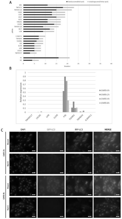
[0031] 1.1 Rat preparation: 4 W SD rats with a weight of about 140 g were purchased from Slack Company and kept in the SPF room of Tongji University Animal Center. Rats were randomly divided into four groups, PBS control group, GMFB treatment group, GMFB-Met treatment group, GMFB-Mutant treatment group.

[0032]1.2 Intravitreal injection: Rats were anesthetized by intraperitoneal injection of 2% pentobarbital sodium (1ml / 400g body weight) before injection, 1× Sumianxin (0.1ml / 200g) for muscle relaxation, and then given a drop of 0.5% tropica Mydriasis with amine (WuxiShanhe Group, Jiangsu, China), topical anesthesia with a drop of 0.4% oxybucaine hydrochloride (Eisai Co Ltd, Tokyo, Japan). Under a stereomicroscope, first use a 1ml syringe to pierce a small hole vertically from the corneal limbus, and then use an injection needle to inject the corresponding liquid into the vitreous cavity from the hole. The injection volume was 8 μl...

Embodiment 2

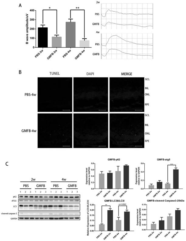
[0034] Electroretinogram ERG detection of physiological function in SD rats

[0035] 2.1 Instruments: APS Automatic Visual Electrophysiology Tester (APS-2000) was purchased from Chongqing Kanghua Technology Co., Ltd.

[0036] Dark adaptation: The day before the visual electrophysiological function test, the SD rats were transferred to a dark room for dark adaptation. Start doing it the next day.

[0037] 2.2 Preparation of rats: intraperitoneal injection of 2% pentobarbital sodium (1ml / 500g body weight) to rats for anesthesia, 1× Sumianxin (0.1ml / 200g) for muscle relaxation, and then give a drop of 0.5% tropica Mydriasis with amine (Wuxi Shanhe Group, Jiangsu, China), topical anesthesia with a drop of 0.4% oxybucaine hydrochloride (Eisai Co Ltd, Tokyo, Japan), and a little conductive paste applied to each eye.

[0038] Insert the electrodes: connect the ground wire to the tail of the rat, connect the negative electrode between the ears of the rat, and connect the positive el...
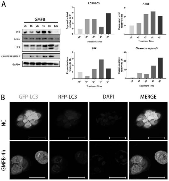
Embodiment 3

[0042] Western-blot detection of apoptosis or autophagy-related protein levels after drug treatment or injection

[0043] 3.1 Protein extraction: Seed the cells in a six-well cell culture dish, after corresponding treatment, add 150 μl of RIPA lysate containing Protease Inhibitor Cocktail to each well, collect the cells into a new EP tube with a cell scraper, and put them on ice Incubate for 30min, centrifuge at 10000rpm at 4°C for 15min, gently aspirate the supernatant into a clean centrifuge tube, store at -80°C for later use.

[0044] 3.2 Determination of protein concentration: BCA quantitative method is used for protein quantification, and the specific method is as follows: prepare a standard protein solution of 2 μg / μl, and store it at -20°C for later use; prepare a working solution according to the number of standards and samples, and use 50 for reagent A and reagent B: The volume ratio of 1 is fully mixed, and it is prepared and used immediately; the total volume of the...


PUM


Abstract

The invention provides an inhibitor for autophagy and apoptosis of retinal pigment epithelial cells RPE and application of the inhibitor, and relates to the technical field of biological medicines. The invention relates to an inhibitor for autophagy and apoptosis of retinal pigment epithelial cells RPE. The inhibitor comprises an antagonist Met of a tumor necrosis factor superfamily receptor FAS.It is verified that the tumor necrosis factor superfamily receptor FAS can serve as a receptor of GMFB to activate downstream autophagy and apoptosis pathways, the antagonist Met of the tumor necrosisfactor superfamily receptor FAS can inhibit activation of the FAS and downstream related pathways of the FAS, cell apoptosis and autophagy reactions are reduced, and therefore the morbidity process of diabetic retinopathy is delayed. The inhibitor can be used for preparing medicines for delaying and / or treating diabetic retinopathy in the early stage of disease.

Description

technical field [0001] The invention belongs to the technical field of biomedicine, and specifically relates to an inhibitor of autophagy and apoptosis of retinal pigment epithelial cells RPE and an application thereof. Background technique [0002] Diabetic retinopathy (DR) is a specific microvascular complication of diabetes. It is chronic, progressive, and potentially harmful to vision. It is the leading cause of blindness in the working population and the elderly. There are more than 93 million DR patients worldwide, of which 17 million suffer from proliferative DR (proliferative DR, PDR), and 28 million suffer from vision-threatening DR (vision threatening DR, VTDR). According to the epidemiological survey in China, the incidence rates of DR and PDR in Chinese diabetic patients are 23% and 2.83%, respectively, and almost half of patients with diabetes for more than 20 years will suffer from DR. According to the severity of the lesion and the formation of new blood vess...

Claims


Application Information

Patent Timeline
Patent Type & Authority Applications(China)
IPC IPC(8): A61K45/06A61K38/18A61P3/10A61P9/10
CPCA61K45/06A61K38/1833A61P3/10A61P9/10
Inventor 吕立夏徐国彤刘彩莹王娟张介平高芙蓉田海滨金彩霞徐晶莹欧庆键
Owner TONGJI UNIV
Why Eureka
Try Eureka