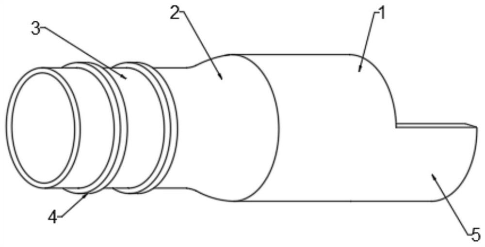
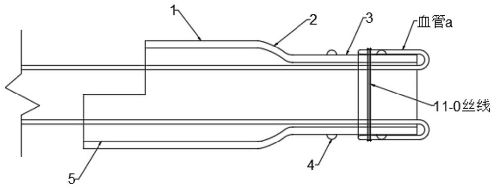
Blood vessel and lymphatic vessel anastomosis sleeve device for microsurgery

A technology of microsurgery and cannula device, applied in medical science, surgery, etc., can solve problems such as limited application, achieve the effect of excellent operation, reduce cost, and improve patency rate

Inactive Publication Date: 2021-02-02
XIANGYA HOSPITAL CENT SOUTH UNIV
View PDF6 Cites 4 Cited by
  • Summary
  • Abstract
  • Description
  • Claims
  • Application Information

AI Technical Summary

Problems solved by technology

If it is used in the field of microsurgery to anastomose small blood vessels and lymphatic vessels (diameter less than 1mm), the application of this cannula device is obviously limited

Method used


Image

  • Blood vessel and lymphatic vessel anastomosis sleeve device for microsurgery
  • Blood vessel and lymphatic vessel anastomosis sleeve device for microsurgery
  • Blood vessel and lymphatic vessel anastomosis sleeve device for microsurgery

Examples


Embodiment 1

[0034] Biocompatibility Test of Polyimide Sleeve

[0035] (1) Cytotoxicity test

[0036] The polyimide material of the present invention is used for cytotoxicity test with L929 mouse fibroblasts, and a negative control group is set.

[0037] The specific method is as follows: sterilize polyimide film with a diameter of 10 cm and a thickness of 5 mm under high temperature and high pressure, and put it into the bottom of a 10 cm petri dish. L929 mouse fibroblasts were cultured in RPMI-1640 medium (containing 10% fetal bovine serum, penicillin 100 U / ml, streptomycin 100 U / ml) in a 5% CO2 incubator at 37°C for 24 hours. Then 0.25% trypsin (containing 0.02% EDTA) was added to digest the monolayer cultured cells, and RPMI-1640 medium containing 10% fetal bovine serum was used to prepare a single cell suspension for electron microscope detection. The same method was used for the negative control group, but there was no polyimide film at the bottom of the petri dish.

[0038] The e...

Embodiment 2

[0044] Animal experiment of small blood vessel anastomosis using polyimide sleeve (in vivo experiment)

[0045] Select 40 healthy male B6 mice, weighing 22-26 g, to construct mouse hindlimb orthotopic transplantation models. 40 mice were randomly divided into 2 groups, 20 in the experimental group and 20 in the control group, 10 as donors and 10 as recipients.

[0046] Specific experimental method: the hind limbs of the donor mice were anesthetized with isoflurane gas, and the hind limbs were stored on ice after harvesting. The recipient mice were anesthetized with isoflurane gas to expose the femoral artery and femoral vein, cut off the hindlimb, connect the donor's hindlimb to the recipient's hindlimb muscles, and fix the femoral bone with intramedullary needles. The core of mouse hindlimb orthotopic transplantation is the anastomosis of the femoral artery (0.8mm in diameter) and the femoral vein (1mm in diameter). The mice in the experimental group were anastomosed end-to-...


PUM


Abstract

The invention discloses a blood vessel and lymphatic vessel anastomosis sleeve device for microsurgery, and belongs to the technical field of microsurgery. The blood vessel and lymphatic vessel anastomosis sleeve device comprises a pipeline a, wherein one end of the pipeline a is connected with a pipeline b through a connecting part; two annular convex parts are integrally arranged on the outer side wall of the pipeline b, and the annular convex parts are used for fixing a blood vessel or a lymphatic vessel; and the outer diameter of the pipeline a is larger than that of the pipeline b, and the inner diameter of the pipeline a is larger than that of the pipeline b. According to the sleeve device, the sleeve device can be used for quickly and efficiently anastomosing blood vessels and lymphatic vessels with calibers of 0.4 mm to 1 mm, two broken-end blood vessels are in well involution (intima-to-intima), the device is not in contact with the inner wall of a lumen and blood flow in thelumen, the patency rate after anastomosis is high, the leakage rate is low, and the occurrence rate of postoperative vascular crisis is low. The device not only can be suitable for blood vessels withthe diameter smaller than 1 mm, but also can be suitable for anastomosis of blood vessels with the diameter larger than 1 mm, fills the application blank of other cannulas, and meanwhile has high universality.

Description

technical field [0001] The invention relates to the technical field of microsurgery, in particular to a cannula device for anastomosis of blood vessels and lymph vessels in microsurgery. Background technique [0002] With the progress and development of modern medicine, the concept of minimally invasive surgery has become the mainstream. Microsurgery is an operation performed with the help of a magnifying microscope. The operation is delicate and causes little trauma to soft tissues. It is widely used in burn plastic surgery, neurosurgery, orthopedics and other departments. [0003] The core of microsurgery lies in the anastomosis of small blood vessels and lymphatic vessels. Although 9-0 sutures and even thinner surgical sutures such as 12-0 are the choice of clinicians for manual anastomosis of blood vessels and lymphatic vessels, manual vascular anastomosis requires long-term animal surgical training and a lot of experience before it can be safely performed It is implem...

Claims


Application Information

Patent Timeline
Patent Type & Authority Applications(China)
IPC IPC(8): A61B17/11
CPCA61B17/11A61B2017/1135
Inventor 贺继强唐举玉吴攀峰卿黎明周征兵俞芳
Owner XIANGYA HOSPITAL CENT SOUTH UNIV
Why Eureka
Try Eureka