Preparation method of biological 3D printing composite ink for repairing cartilage defects

A 3D printing and cartilage technology, applied in the field of biological 3D printing, can solve the problems of natural three-dimensional microenvironment that cannot simulate cell growth, cell adhesion, unsatisfactory directional differentiation, and difficulty in accurate imitation, and achieve good clinical application prospects and value, rapid cross-linking and curing, and high biocompatibility

Inactive Publication Date: 2021-02-02
SECOND MILITARY MEDICAL UNIV OF THE PEOPLES LIBERATION ARMY +1
View PDF5 Cites 11 Cited by
  • Summary
  • Abstract
  • Description
  • Claims
  • Application Information

AI Technical Summary

Problems solved by technology

[0004] Aiming at the deficiencies of the prior art, the present invention provides a preparation method and application of a bio-3D printing composite ink containing cartilage extracellular matrix, which solves the problem that the current bio-3D printing ink proposed in the background technology simply uses alginate, Hydrogels prepared from chitosan, methacryloyl gelatin, hyaluronic acid or collagen, etc., cannot simulate the natural three-dimensional microenvironment of cell growth due to their biological inertness. The performance is not satisfactory, and even some raw materials will produce components that are toxic to cells after degradation, and so far, no materials and inks that can accurately mimic the natural extracellular matrix have been found, because the complex 3D structure of native ECM and material composition make it very difficult to accurately simulate the problem of

Method used


Image

  • Preparation method of biological 3D printing composite ink for repairing cartilage defects
  • Preparation method of biological 3D printing composite ink for repairing cartilage defects
  • Preparation method of biological 3D printing composite ink for repairing cartilage defects

Examples


Embodiment 1

[0032] Embodiment 1. The present invention provides a technical solution: a method for preparing a bio-3D printing composite ink for repairing cartilage defects, comprising the following steps:

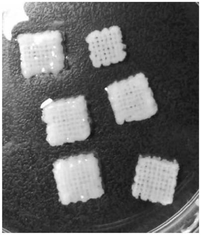
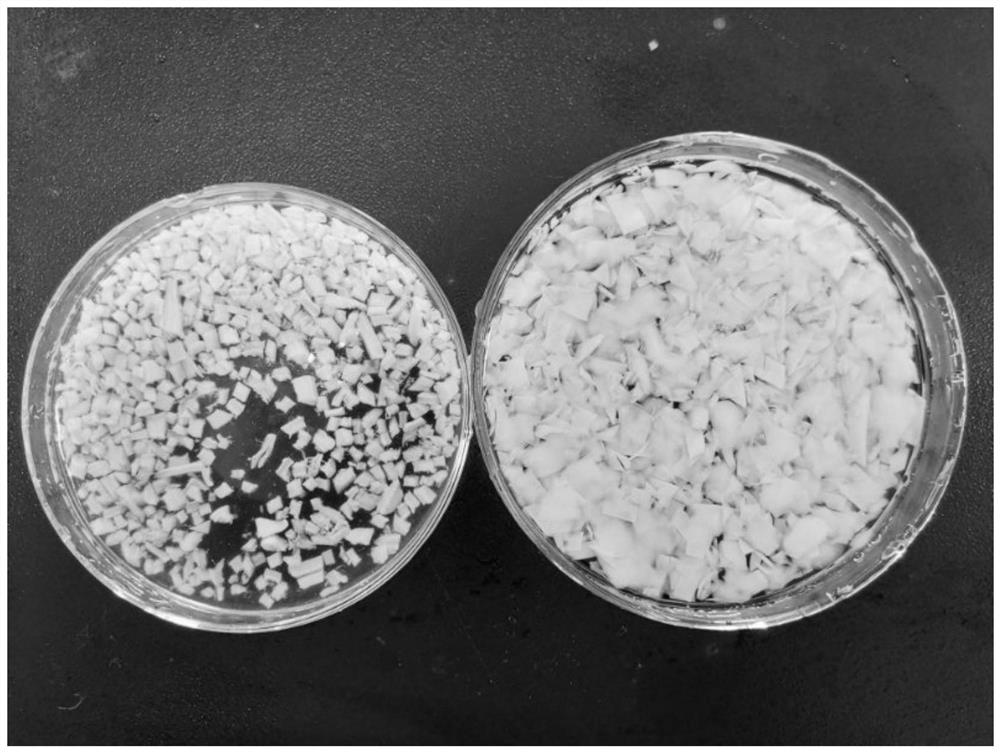
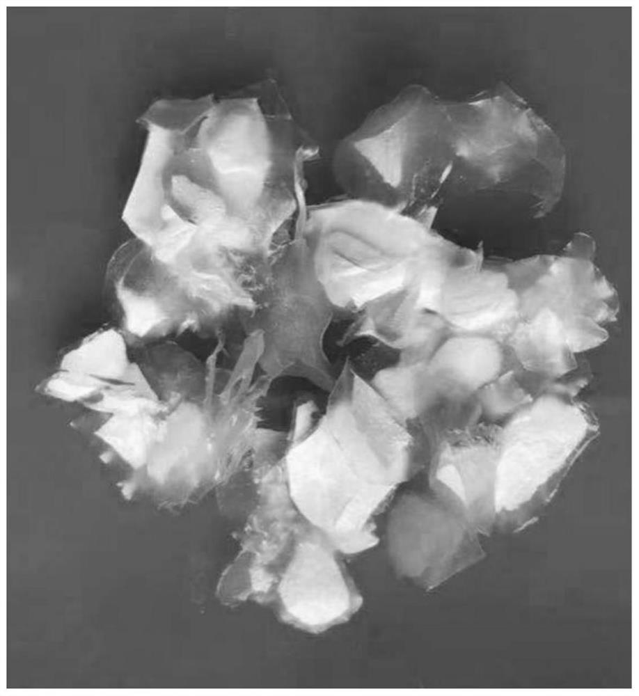
[0033] S1. Preparation of cartilage granules: take the fresh articular surface hyaline cartilage or costal cartilage of animals, cut off the hyaline cartilage from the articular surface with a sharp scalpel blade to obtain cartilage slices, and then cut the cartilage slices into granules with a particle size of 1-2mm 3 (Such as figure 1 shown);

[0034] S2. Decellularization treatment: Soak cartilage particles in hypotonic Tris-HCl buffer, the concentration of hypotonic Tris-HCl buffer is 10mM, PH=8, and then cycle freeze-thaw at -80°C ~ 37°C 6 cycles; then digest with 0.25% trypsin at 37°C for 24 hours, and replace the trypsin solution every 4 hours; then wash away the trypsin with hypertonic Tris-HCl buffer solution, The permeated Tris-HCl buffer solution is composed of NaCl 1.5M...

Embodiment 2

[0041] Embodiment 2. The present invention provides a technical solution: a method for preparing a bio-3D printing composite ink for repairing cartilage defects, comprising the following steps:

[0042] S1. Preparation of cartilage granules: take the fresh articular surface hyaline cartilage or costal cartilage of animals, cut off the hyaline cartilage from the articular surface with a sharp scalpel blade to obtain cartilage slices, and then cut the cartilage slices into granules with a particle size of 1-2mm 3 (Such as figure 1 shown);

[0043] S2. Decellularization treatment: Soak cartilage particles in hypotonic Tris-HCl buffer, the concentration of hypotonic Tris-HCl buffer is 10mM, PH=8, and then cycle freeze-thaw at -80°C ~ 37°C 6 cycles; then digest with 0.25% trypsin at 37°C for 24 hours, and replace the trypsin solution every 4 hours; then wash away the trypsin with hypertonic Tris-HCl buffer solution, The permeated Tris-HCl buffer solution is composed of NaCl 1.5M...

Embodiment 3

[0050] Embodiment 3. The present invention provides a technical solution: a method for preparing a bio-3D printing composite ink for repairing cartilage defects, comprising the following steps:

[0051] S1. Preparation of cartilage granules: take the fresh articular surface hyaline cartilage or costal cartilage of animals, cut off the hyaline cartilage from the articular surface with a sharp scalpel blade to obtain cartilage slices, and then cut the cartilage slices into granules with a particle size of 1-2mm 3 (Such as figure 1 shown);

[0052] S2. Decellularization treatment: Soak cartilage particles in hypotonic Tris-HCl buffer, the concentration of hypotonic Tris-HCl buffer is 10mM, PH=8, and then cycle freeze-thaw at -80°C ~ 37°C 6 cycles; then digest with 0.25% trypsin at 37°C for 24 hours, and replace the trypsin solution every 4 hours; then wash away the trypsin with hypertonic Tris-HCl buffer solution, The permeated Tris-HCl buffer solution is composed of NaCl 1.5M...


PUM


Abstract

The invention discloses a preparation method of biological 3D printing composite ink for repairing cartilage defects, and relates to the technical field of biological 3D printing. The preparation method of the biological 3D printing composite ink for repairing the cartilage defects comprises the following steps: obtaining fresh transparent articular facet cartilage or costal cartilage from animals, removing cartilage cells by adopting physical, chemical and biological methods, and carrying out sterilizing and freeze-drying; carrying out grinding under the condition of low temperature with liquid nitrogen so as to obtain extracellular matrix powder; and then, uniformly mixing the extracellular matrix powder with a mixed material according to a certain ratio, adding deionized water, and allowing dispersing and dissolving so as to form a uniform and stable suspension, namely a target product. The preparation method of the biological 3D printing composite ink for repairing the cartilage defects provided by the invention has the following beneficial effects: the obtained extracellular matrix powder can be combined with different materials so as to satisfy biological 3D printing composite ink meeting different requirements; and moreover, the ink can be rapidly crosslinked and cured, and is relatively low in cytotoxicity, relatively high in biocompatibility and capable of being co-printed with stem cells so as to meet tissue engineering repair requirements. Thus, the ink has relatively good clinical application prospects and values.

Description

technical field [0001] The invention relates to the technical field of biological 3D printing, in particular to a preparation method of biological 3D printing composite ink for repairing cartilage defects. Background technique [0002] Bioconstruction is an emerging research field, which generally refers to the process of creating biological tissues with a specific hierarchical structure by means of bioengineering. Traditional bioconstruction techniques commonly include particle leaching, freeze-drying, and electrospinning, etc. Although these technologies can Generate a series of biological materials with 3D structure, but often have poor reproducibility and functional scalability, 3D bioprinting has become a new biological construction method, based on digitally controlled automatic deposition technology, can be used for tissue structure The construction process provides precise control, high repeatability and functional scalability. Bio-ink is an ink that can be used in b...

Claims


Application Information

Patent Timeline
Patent Type & Authority Applications(China)
IPC IPC(8): A61L27/18A61L27/20A61L27/22A61L27/24A61L27/36A61L27/50B33Y70/10B33Y80/00
CPCA61L27/18A61L27/20A61L27/222A61L27/24A61L27/3633A61L27/3687A61L27/3691A61L27/50A61L2430/06B33Y70/00B33Y80/00C08L5/04C08L5/08C08L67/04
Inventor 吴俊钱齐荣邓建华周义钦韩亚光
Owner SECOND MILITARY MEDICAL UNIV OF THE PEOPLES LIBERATION ARMY
Why Eureka
Try Eureka