Detection method of lipid content in biological liver tissue

A technology of liver tissue and detection method, which is applied to measurement devices, instruments, scientific instruments, etc., can solve the problems of poor repeatability, low sensitivity, interference in the detection process of lipid content, etc., and achieves improvement of lipid extraction rate and accuracy. , Taking into account the effect of lipid extraction rate and impurity removal rate

Active Publication Date: 2021-06-04
THE AFFILIATED HOSPITAL OF QINGDAO UNIV
View PDF12 Cites 0 Cited by
  • Summary
  • Abstract
  • Description
  • Claims
  • Application Information

AI Technical Summary

Problems solved by technology

However, when using the method provided by this patent to extract lipids, the interface between the upper organic phase and the lower aqueous phase is unstable, which may easily cause the problems of low lipid extraction rate and poor reproducibility, and the detection sensitivity is relatively low. Low; moreover, the lipid extracted by this method will contain more polar matrix, which will interfere with the subsequent detection process of lipid content, and the detection result of lipid content in liver tissue is not accurate enough

Method used


Image

  • Detection method of lipid content in biological liver tissue
  • Detection method of lipid content in biological liver tissue
  • Detection method of lipid content in biological liver tissue

Examples


preparation example Construction

[0041] In step S3, the preparation method of the filler includes the following steps:

[0042] S31, adding silica gel particles into the hydrochloric acid solution, fully mixing at 90-100° C. for 14-16 hours, filtering, washing and drying to obtain activated silica gel;

[0043] S32. Add methylimidazole and 3-chloropropyltriethoxysilane into the acetonitrile solution, react at 100-110°C for 16-20 hours, then add potassium hexafluorophosphate, continue the reaction for 24-28 hours, filter and Collect the filtrate;

[0044] S33. After mixing the filtrate obtained in step S32 with toluene, add the activated silica gel obtained in step S31, react at 100-110°C for 22-26 hours, filter, wash and dry the product to obtain functionalization silica gel;

[0045] S34. Add the functionalized silica gel obtained in step S33 into the acetone solution of silver tetrafluoroborate, mix thoroughly for 8-10 hours under the condition of avoiding light, filter, wash and dry to obtain silica gel ...

Embodiment 1

[0051] This embodiment provides a method for detecting lipid content in biological liver tissue, comprising the following steps:

[0052] S1. Homogenization and primary extraction

[0053] Add 100mg of mouse liver tissue into the homogenization tube containing ceramic microbeads, then add 5mL of the first mixed solvent made of isopropanol and acetonitrile at a volume ratio of 1:2.5, and homogenize at a speed of 3000r / min for 15s , and the homogenate was obtained after repeating the homogenization twice.

[0054] After continuing to centrifuge the homogenate at a speed of 3000 r / min for 10 min, take the supernatant as the first extract.

[0055] S2, secondary extraction

[0056] Take 500 μL of the first extract obtained in step S1, add 750 μL of the second mixed solvent formed by mixing n-hexane and ethyl acetate at a volume ratio of 3.5:1, and then add 750 μL of 1.5% aqueous acetic acid solution, After vortexing for 20 s, centrifuge at 3000 r / min for 10 min, and take the su...

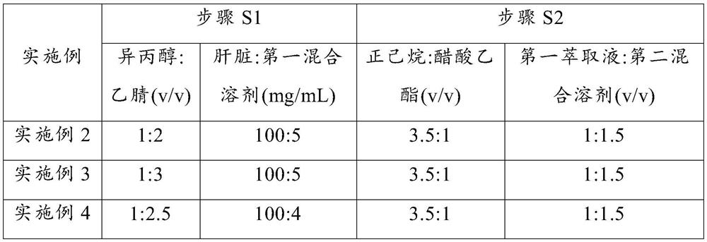
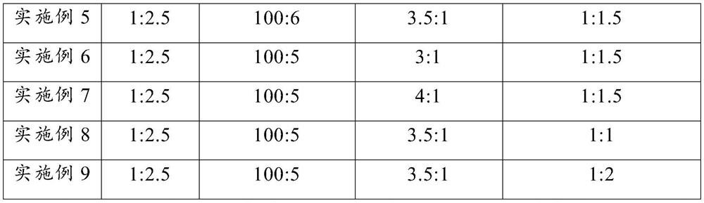
Embodiment 2~9 and comparative example 1~2

[0074] Embodiments 2 to 9 respectively provide a detection method for the lipid content of biological liver tissue. Compared with embodiment 1, the difference is that some parameters in steps S1 to S2 are changed. The detection parameters corresponding to each embodiment are as follows: As shown in Table 2, the rest of the steps are consistent with those in Example 1, and will not be repeated here.

[0075] The corresponding parameter of each step in the embodiment 2~9 of table 2

[0076]

[0077]

[0078] Comparative Example 1 and Comparative Example 2 respectively provide a detection method for the lipid content of biological liver tissue. Compared with Example 1, the difference is that in step S1 of Comparative Example 1, deionized water is used instead of the first mixed solvent to carry out Detection; step S2 was not performed in comparative example 2, and the first extract obtained in step S1 was directly subjected to solid-phase extraction; the rest of the steps i...


PUM


Abstract

The invention provides a method for detecting lipid content in biological liver tissue. In the present invention, the first mixed solvent is added to the biological liver tissue, and after homogenization and centrifugation, the first extract is obtained; the second mixed solvent is added to the first extract, and an aqueous acetic acid solution is added to obtain the The second extraction solution; then the silica gel that is loaded with silver ions is used as a filler, and the second extraction solution is carried out to solid-phase extraction by using a solid-phase extraction column containing the filler to obtain a lipid extract, and the chromatographic-mass spectrometry technology is used to extract the It detects and obtains the contents of various lipids in the biological liver tissue. Through the above method, the present invention can fully extract various lipids contained in biological liver tissue through two-step liquid phase extraction, and perform solid phase extraction on the extract by preparing silica gel loaded with silver ions, so as to realize effective enrichment of lipids. The set can effectively improve the sensitivity and accuracy of lipid content detection, and has a good application prospect.

Description

technical field [0001] The invention relates to the technical field of lipid content detection, in particular to a method for detecting lipid content of biological liver tissue. Background technique [0002] Lipids are generally defined as a class of small molecular compounds that are insoluble in water and easily soluble in organic solvents in nature, and are very important substances in living organisms. As the main components of biological membranes, lipids have molecular diversity and various biological functions, including providing a suitable environment for protein interaction, storing energy, and serving as important messenger molecules. In various organs of the organism, the liver plays an important role in the digestion, absorption, synthesis, decomposition and transportation of lipids. Accurate and effective detection of lipid content in biological liver tissue is of great importance for liver lipid metabolomics. research is of great significance. [0003] At pr...

Claims


Application Information

Patent Timeline
Patent Type & Authority Patents(China)
IPC IPC(8): G01N30/02G01N30/06G01N30/08G01N30/72G01N30/86
CPCG01N30/02G01N30/06G01N30/08G01N30/7206G01N30/8675G01N2030/062
Inventor 曲林林杨振全
Owner THE AFFILIATED HOSPITAL OF QINGDAO UNIV
Why Eureka
Try Eureka