Composite osteochondral scaffold as well as preparation method and application thereof

An osteochondral and cartilage layer technology, applied in the field of 3D printing materials, can solve the problems of single biological scaffold, single material structure, and differences in mechanical and biological properties.

Active Publication Date: 2021-06-25
SHANGHAI UNIV +1
View PDF3 Cites 6 Cited by
  • Summary
  • Abstract
  • Description
  • Claims
  • Application Information

AI Technical Summary

Problems solved by technology

Most of the existing bioscaffolds printed by 3D are single material and single structure
However, cartilage and subchondral bone have different structures, and there are large differences in their mechanical and biological properties. Traditional scaffolds with a single material and a single structure are difficult to meet the repair requirements

Method used


Image

  • Composite osteochondral scaffold as well as preparation method and application thereof
  • Composite osteochondral scaffold as well as preparation method and application thereof
  • Composite osteochondral scaffold as well as preparation method and application thereof

Examples


preparation example Construction

[0042] The present invention also provides a method for preparing the composite osteochondral scaffold described in the above technical solution, comprising the following steps:

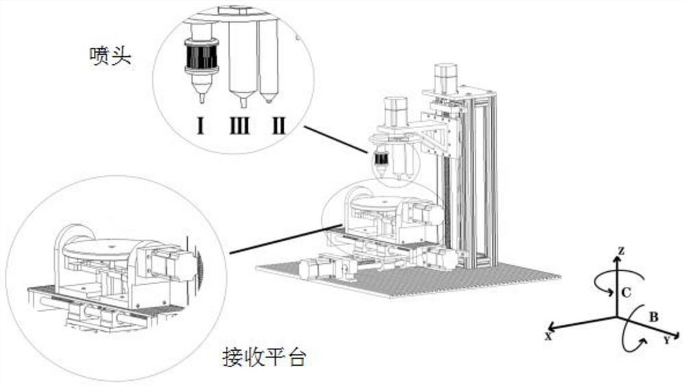
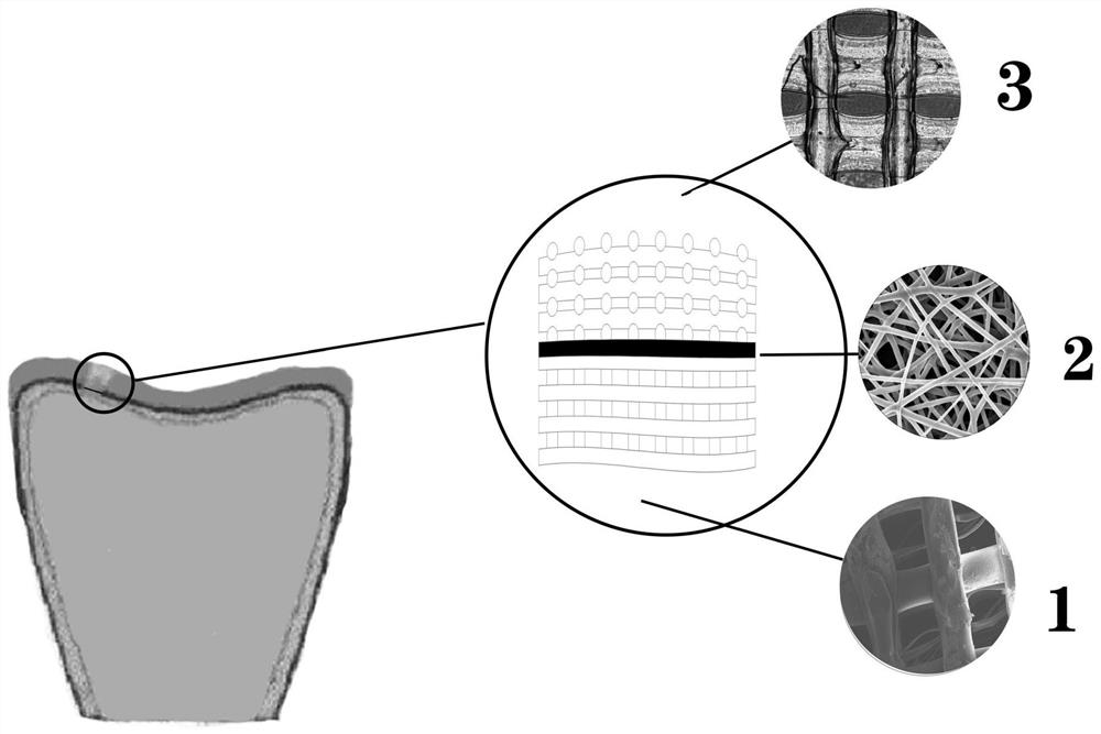
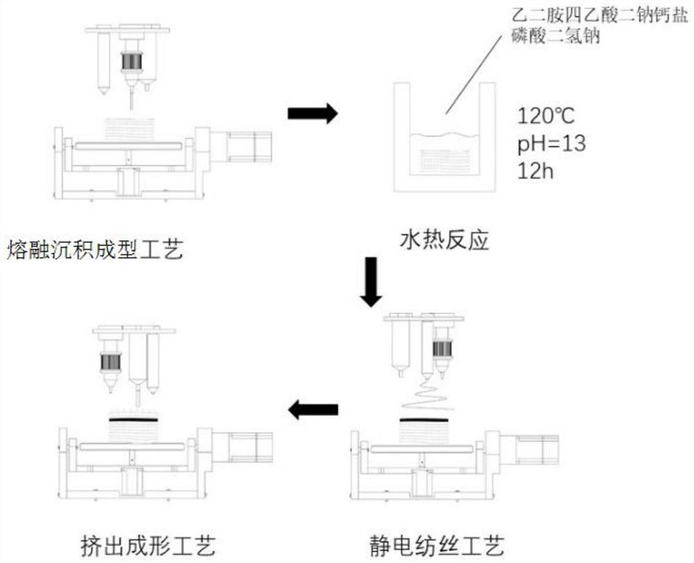
[0043] Using fused deposition modeling process to 3D print polyetheretherketone into subchondral bone layer;

[0044] Coating a biocompatible solution on the surface of the subchondral bone layer by a hydrothermal method to form a biocompatible layer;

[0045] Using an electrospinning process to 3D print a fibrous film-like transition layer on the surface of the biocompatible layer;

[0046] A cartilage layer with a network structure is 3D printed on the surface of the transition layer by an extrusion molding process, and the extruded raw material contains cartilage matrix and biological factors, and the cartilage matrix is ​​a hydrogel material.

[0047] The invention utilizes a fused deposition modeling process to 3D print polyetheretherketone into a subchondral bone layer. In the present inventi...

Embodiment 1

[0073] CT scans the point cloud data of the articular cartilage defect, converts the point cloud data obtained from the CT scan into a solid model through reverse design, and builds a three-dimensional model of the defect with reference to the complete cartilage tissue structure, and adds boundary conditions to the obtained model, and conducts limited Meta-analysis to obtain its stress-strain distribution; according to the stress-strain distribution, the path planning for 3D printing of the subchondral bone model is carried out;

[0074] Nozzle I (nozzle diameter: 0.4mm) is loaded with polyether ether ketone, and the nozzle I moves in the three directions of x, y, and z, and cooperates with the rotation of the B-axis and C-axis of the receiving platform to print according to the planned printing path (the temperature of the fusion deposition 395°C) subchondral bone layer to obtain a reticular subchondral bone layer with a pore size of 800 μm;

[0075] Dissolve 3.75g of ethylened...

Embodiment 2

[0079] The composite osteochondral scaffold was prepared according to the method of Example 1, except that the temperature of the hydrothermal reaction was 180°C.


PUM

PropertyMeasurementUnit
thicknessaaaaaaaaaa
porosityaaaaaaaaaa
pore sizeaaaaaaaaaa

Abstract

The invention belongs to the technical field of 3D printing materials, and particularly relates to a composite osteochondral stent as well as a preparation method and application thereof. The invention provides a composite bone cartilage scaffold. The composite bone cartilage scaffold sequentially comprises a subchondral bone layer, a fibrous membrane-shaped transition layer and a cartilage layer with a net structure from bottom to top; the subchondral bone layer is made of polyether-ether-ketone, and the surface of the subchondral bone layer is coated with a biocompatible layer; the material of the cartilage layer comprises a cartilage matrix and biological factors, and the cartilage matrix is a hydrogel material; and the cartilage layer contains biological factors. Biological factors contained in the cartilage layer are beneficial to the growth of cartilage; The transition layer can realize the exchange of nutrient substances between the cartilage layer and the subchondral bone layer, and meanwhile, the cartilage layer and the subchondral bone layer have relatively independent development environments, so that the problem of fibrosis is avoided. The composite osteochondral scaffold provided by the invention can be well integrated with surrounding tissues, and integrated repair of cartilage and subchondral bone can be realized.

Description

technical field [0001] The invention belongs to the technical field of 3D printing materials, and in particular relates to a composite osteochondral scaffold and its preparation method and application. Background technique [0002] Articular cartilage lesions are a common clinical disease, most of which are caused by trauma, tumors, accidents, etc. Because cartilage tissue lacks blood vessels, nerves, and undifferentiated cells, its ability to repair itself is limited. From the perspective of anatomical structure, cartilage and subchondral bone are closely connected. Usually, most articular cartilage lesions will affect the subchondral bone, and cartilage and subchondral bone affect each other in terms of biological functions. Therefore, when performing tissue repair, it is necessary to Consider both at the same time. [0003] Currently commonly used solutions include microfracture, autologous cartilage transplantation, and allogeneic cartilage transplantation. Among them...

Claims


Application Information

Patent Timeline
Patent Type & Authority Applications(China)
IPC IPC(8): A61L27/18A61L27/50A61L27/52A61L27/54A61L27/36B33Y70/10B33Y80/00B33Y10/00A61F2/28
CPCA61L27/18A61L27/50A61L27/52A61L27/54A61L27/3654A61L27/3612B33Y70/10B33Y80/00B33Y10/00A61F2/28A61L2430/06A61L2400/12A61L2300/414C08L61/16
Inventor 胡庆夕王亚豪王国印张海光
Owner SHANGHAI UNIV
Why Eureka
Try Eureka