
Virtual probe for remote cardiac ultrasound and diagnosis and treatment system

A technology of virtual probe and diagnosis and treatment system, applied in ultrasonic/sonic/infrasonic diagnosis, ultrasonic/sonic/infrasonic Permian technology, sound wave diagnosis, etc. It can solve the problem that the virtual probe is not small enough to be integrated, the ultrasonic image of the patient is accidentally injured, and the data exchange is not enough Real-time and other issues to achieve the effect of saving business training time, avoiding accidents in time, and clear ultrasound imaging

Pending Publication Date: 2021-07-30
SHANDONG UNIV
View PDF12 Cites 0 Cited by
  • Summary
  • Abstract
  • Description
  • Claims
  • Application Information

AI Technical Summary

Problems solved by technology

However, there are problems with the virtual probe and the remote ultrasonic inspection method proposed in the current remote ultrasound patent: (1) There is no force feedback device, so experts cannot remotely control the force exerted by the robotic arm, which may easily cause excessive force to accidentally injure the patient or The ultrasonic image is blurred due to too small force; (2) The virtual probe is not small enough to be integrated, and cannot map all the operation actions of the expert, and additional sensors or keyboard and mouse are needed to control some degrees of freedom; (3) Ultrasound scanning for The parts are fixed and simple parts such as the liver and lungs, and there is no remote ultrasonic scanning device for complicated organs such as the heart; (4) There is a delay in data transmission, and real-time reproduction cannot be achieved
[0006] After retrieval, the Chinese invention patent with the publication number CN110993087A uses a gyroscope to collect attitude information, but needs to place an additional infrared sensor outside the probe to collect position information, so it is not integrated enough, and an additional sensor is used; at the same time, the data uses the TCP / IP protocol Transmission and data exchange are not real-time enough; the third problem is that the product puts the probe on a plane, which is close to the operation habits of experts for abdominal ultrasound exploration, but it is not suitable for cardiac ultrasound exploration
During echocardiography, because there are many bony structures in the chest wall, the contour of the chest wall is more undulating. At the same time, the patient is placed in a lateral position during the ultrasound examination. Therefore, the Chinese patent invention with the publication number CN110993087A is only applicable to the detection of flat and Fixed and simple ultrasonic exploration, not suitable for complex ultrasonic exploration such as the heart

Method used


Image

  • Virtual probe for remote cardiac ultrasound and diagnosis and treatment system
  • Virtual probe for remote cardiac ultrasound and diagnosis and treatment system
  • Virtual probe for remote cardiac ultrasound and diagnosis and treatment system

Examples


Embodiment 1

[0051] The base of the robotic arm is fixed, and the patient's movement is the main way to adjust the position.

[0052] The specific content is as follows:

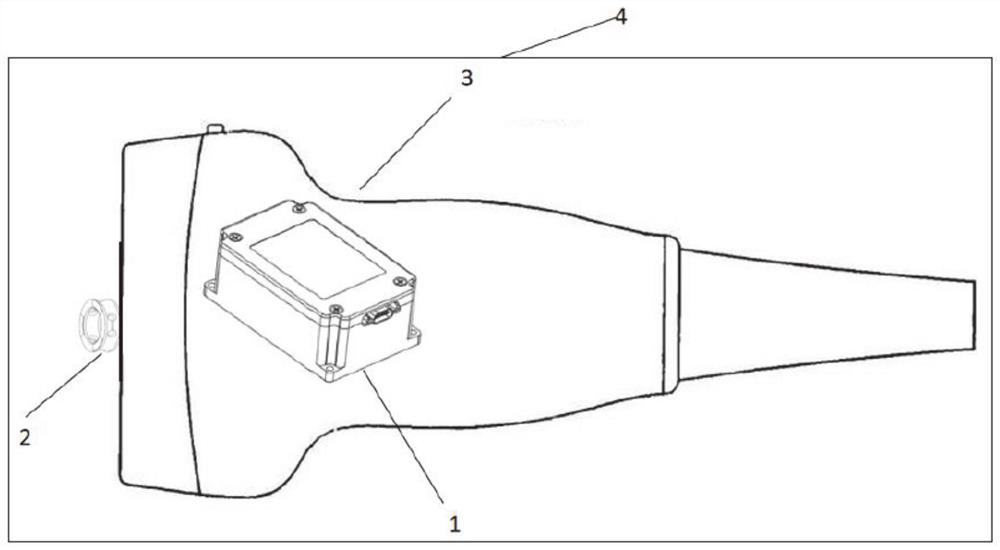
[0053] Such as figure 1 As shown, the virtual ultrasonic probe 1 is used to transmit the position and posture of the remote-end expert when operating the probe and the force acting on the human body model to the on-site mechanical arm to reproduce the expert's actions.

[0054] In a preferred embodiment, the virtual ultrasonic probe consists of a pose sensor 1 , a force sensor 2 and a 3D printed shell 3 .

[0055] The pose sensor 1 is fixed inside the 3D printed shell 3, and outputs the position and direction of the probe in real time.

[0056] In a preferred embodiment, the pose sensor 1 is installed on the center of gravity of the virtual probe 4, and the angular velocity output by the gyroscope is integrated to obtain the Euler angle. In order to avoid the phenomenon of gimbal lock in the Euler angle, the Euler angl...

Embodiment 2

[0071] The field end uses a mobile robot platform. The difference from Example 1 is that the base of the manipulator in Example 1 is fixed, so the working space of the manipulator is limited, and the manipulator in some detection postures cannot reach it. Therefore, the patient is required to Adjust your posture according to the instructions of the on-site staff, so that your part to be checked is always in the working space of the robotic arm. This optimal case uses the patient's movement as the main position adjustment method, which is suitable for the premise that the patient is in good physical condition and can move autonomously Down.

[0072] In this embodiment, the fixed robotic arm is replaced by a movable robot platform. The robot can move freely while holding the ultrasonic probe, and is no longer bound by the detection position. The position and direction of the probe should be adjusted until the posture is suitable, and then the posture and posture of the probe sho...

Embodiment 3

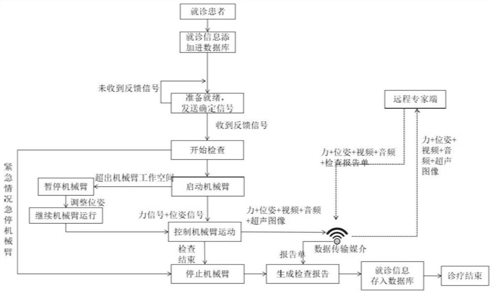
[0083] Figure 4 The direction of signal flow between the various components of teleultrasonography is given. The flow direction of the signal and the connection relationship between the various components are explained in detail below:

[0084] Remote ultrasonic examination is divided into remote end and on-site end. The equipment of each part is composed of: the remote end is composed of ultrasound operation experts, video equipment, virtual ultrasound probe, imitation human body model, ultrasound image display equipment, and server. The on-site end consists of on-site staff, patients, robot platforms, treatment beds, video equipment, servers, ultrasound probes, ultrasound scanners, and ultrasound image display equipment.

[0085] Specifically, the output end of the pose sensor is connected to the computer through a serial port. In order to avoid the gimbal lock phenomenon that is prone to appear in the Euler angle, the pose Euler angle collected by the pose sensor is conv...


PUM


Abstract

The invention provides a virtual probe for remote cardiac ultrasound and a diagnosis and treatment system. The virtual probe comprises a shell, a pose detection element and a force detection element; the pose detection element is fixed in the shell, and the force detection element is fixed on a detection surface, used for being in contact with the bionic model, of the shell. The pose detection element and the force detection element are both in communication connection with the first control terminal, the first control terminal is in communication connection with the second control terminal, and the audio and video acquisition module and the robot control terminal are both in communication connection with the second control terminal; the first control terminal generates a robot mechanical arm control instruction according to the received virtual probe pose and force applied to the bionic model and sends the robot mechanical arm control instruction to the second control terminal; the second control terminal controls a mechanical arm of the robot according to the received control instruction so as to control the action of a real probe, and transmits on-site audio and video data and ultrasonic images to the first control terminal in real time. According to the disclosure, the accuracy and timeliness of remote diagnosis and treatment are greatly improved.

Description

technical field [0001] The present disclosure relates to the technical field of remote medical equipment, in particular to a virtual probe and diagnosis and treatment system for remote cardiac ultrasound. Background technique [0002] The statements in this section merely provide background information related to the present disclosure and may not necessarily constitute prior art. [0003] Ultrasound imaging has great advantages over other medical imaging methods due to its non-invasive, instant diagnosis, real-time, and cost-effective advantages. [0004] In the process of ultrasound acquisition, too small pressure will damage the good acoustic coupling and thus affect the quality of ultrasound imaging, while too high pressure will harm the patient and even deform the observed organs. Feedback is used to adjust the probe pose and the contact force between the probe and the patient. The quality of ultrasound images depends on intrinsic factors such as hardware design and di...

Claims


Application Information

Patent Timeline
Patent Type & Authority Applications(China)
IPC IPC(8): A61B8/08A61B8/00G16H40/67
CPCA61B8/0883A61B8/4444A61B8/565A61B8/54G16H40/67
Inventor 刘治张德彩张鹏飞曹艳坤
Owner SHANDONG UNIV
Learn More