Improvements in variant detection

A variant and sample technology, applied in the field of detection of variant DNA, can solve problems such as impossibility, difficulty in monitoring, and possibility of limiting applications

Pending Publication Date: 2021-08-27
CANCER RES TECH LTD
View PDF0 Cites 0 Cited by
  • Summary
  • Abstract
  • Description
  • Claims
  • Application Information

AI Technical Summary

Problems solved by technology

[0006] Although the detection of variants, such as ctDNA, in cell-free DNA-containing samples shows promise in the field of cancer care, there exists a lack of methods and systems for maximizing the signal-to-noise ratio in the presence of low variant (eg, ctDNA) fractions. unmet needs
Furthermo

Method used


Image

  • Improvements in variant detection
  • Improvements in variant detection
  • Improvements in variant detection

Examples


Embodiment 1

[0297] Example 1 - Identification of Patient-Specific Mutations from Tumor and Plasma

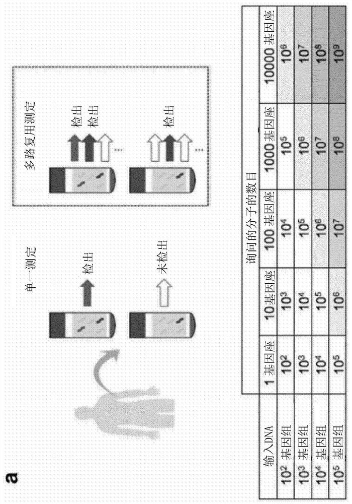
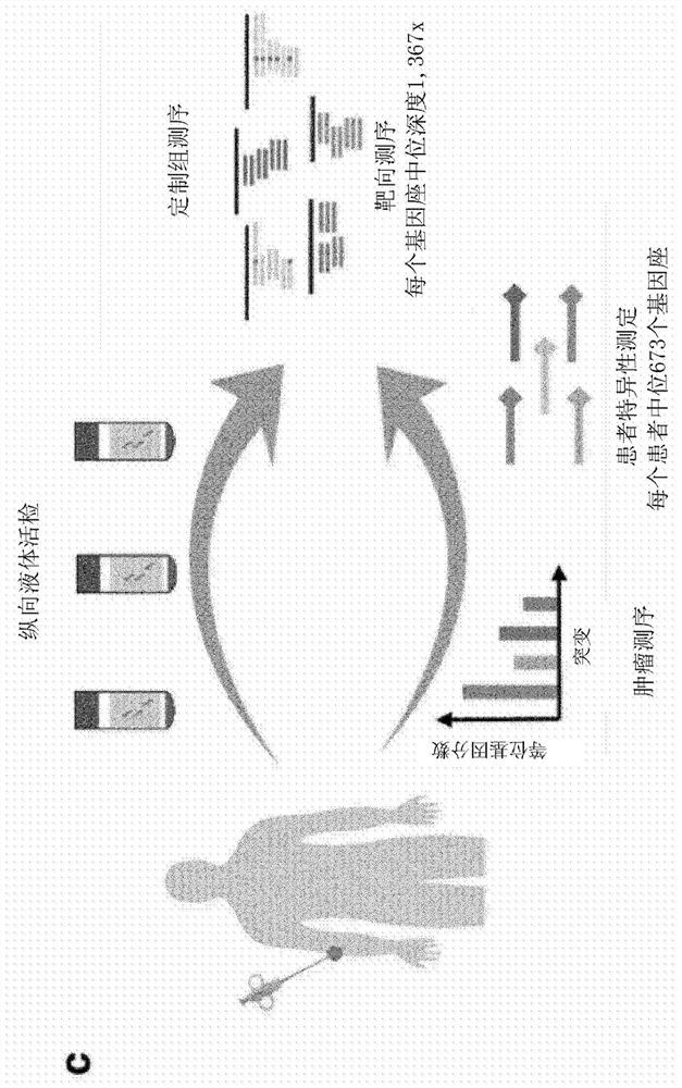
[0298] To obtain high sequencing depth at defined loci mutated in patient tumors, custom hybrid capture sequencing panels were designed based on single nucleotide variants (SNVs) identified in the sequencing of tumor biopsies. SNVs with ≥1 mutant read and ≥10 total reads were selected from exome sequencing (9 patients) or targeted sequencing (1 patient) of baseline metastatic biopsies. The median number of SNVs identified per patient was 673 (IQR 250-1,209; Figure 7a). Patient-specific variants were identified (not shown). Furthermore, to allow de novo identification of mutations in plasma, the coding sequences and untranslated regions of the following genes were included in the panel design: ARID2, BRAF, CDKN2A, NF1, PTEN, and TP53, as well as 37 additional genes commonly mutated in melanoma. Hotspot loci (not shown) among the genes of . The final group design covers 1.527Mbp.

[029...

Embodiment 2

[0301] Example 2 - Characterization of background error rate

[0302] We sought to understand the background error rate (i.e., the rate at which unanticipated mutated bases were observed) with and without error suppression in TAPAS sequencing data. Bases on either side of the patient-specific variants were investigated because they have comparable sequencing depth and are subject to the same technical bias. To take advantage of this off-target sequencing of patient samples, germline events and potential biosignatures were excluded if they occurred multiple times in samples from the same individual (Methods); these loci were left for subsequent de novo mutation calling.

[0303] Error suppression can be achieved by using read compression to determine consensus sequences for entire read families. To achieve this, repeat reads are based on start and end fragment positions (formerly known as 'endogenous barcodes' 11,12 ) and molecular barcodes are grouped into 'read families'. ...

Embodiment 3

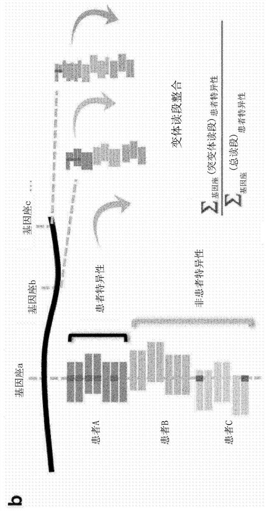
[0304] Example 3 - Variant Read Integration (INVAR)

[0305] Using a strict level of error suppression (consensus sequence required in 90% of family members, minimum family size of 5) and a median of 4.4 ng of input, we obtained a median of 3.2 × 10 5 read family (IQR 8.7×10 4 to 6.2×10 5 ), each covering the loci mutated in that patient's cancer. Under the assumption that each such read family corresponds to a single molecule, we are thus able to probe thousands of target molecules per sample even if the starting material contains only ~1300 genome copies.

[0306] When ctDNA levels are low, many patient-specific loci will have no mutant DNA fragments at that position ( Figure 7 b). Therefore, to overcome sampling error, all patient-specific read families were aggregated and analyzed together using INVAR ( figure 1 b). For each sample, the "global" mutant allele fraction across all patient-specific loci was calculated as follows:

[0307]

[0308] The significance ...


PUM


Abstract

The present invention provides a method for detecting variant cell-free DNA (cfDNA) in a sample obtained from a subject, where analysis of the sample includes a size-selection step which separates out different fragment sizes of DNA. The sample may be a limited volume sample such as a blood, serum or plasma sample of less than 500ul (e.g. a blood or plasma sample of about 50ul), or other sample that has a low content of cfDNA. The sample may have been stored and/or dried and not have been processed to remove cells or cellular material prior to storage. The size-selection step may comprise filtering-out, depleting or removing genomic DNA (gDNA) fragments of > 200 bp, > 300 bp, > 500 bp, > 700 bp, > 1000 bp, > 1200 bp, > 1500 bp, or > 2000 bp prior to analysis, e.g. prior to DNA sequencing. The method may further comprise performing an analysis that summarises or combines data across multiple loci.

Description

technical field [0001] The present invention relates in part to methods for detecting the presence of variant DNA (e.g. circulating tumor DNA (ctDNA)) from sources such as cell-free DNA (cfDNA) such as plasma or for use in forensic Methods to detect variant DNA in applications, pathogen identification, agriculture and environmental species contamination monitoring. In particular, the methods of the invention are useful in the diagnosis, treatment and especially monitoring of cancer, including monitoring following tumor resection. Background technique [0002] Cell-free DNA (cfDNA), such as circulating tumor DNA (ctDNA), is increasingly used as a non-invasive tool to monitor disease burden, response to therapy, and risk of recurrence 1,2 . Following treatment, patients may have low ctDNA levels, and even in advanced disease, concentrations may be below a few copies per sample volume 3 . In this case, due to sampling statistics, a single sample may contain less than one de...

Claims


Application Information

Patent Timeline
IPC IPC(8): C12Q1/6806
CPCC12Q1/6806C12Q2563/149C12N15/1034
Inventor 卡特林·海德尔乔纳森·万尼灿·罗森菲尔德
Owner CANCER RES TECH LTD
Why Eureka
Try Eureka