Corneal neovascularization/lymphatic vessel generation injury model and construction method and application thereof

A corneal neovascularization and damage model technology, which can be used in pharmaceutical formulations, veterinary surgery, veterinary instruments, etc., to solve problems such as increased, unfavorable corneal neovascularization or corneal lymphatic vessels, and scarring.

Pending Publication Date: 2021-10-22
GENERAL HOSPITAL OF PLA
View PDF0 Cites 0 Cited by
  • Summary
  • Abstract
  • Description
  • Claims
  • Application Information

AI Technical Summary

Problems solved by technology

[0006] However, in the prior art, these physical methods induce mouse corneal neovascularization models, and there are certain technical problems
For example, the burn intensity of the thermal burn model is difficult to control. When the intensity is too high, it is easy to cause corneal perforation and scar formation, resulting in the failure of modeling; the chemical burn model is also difficult to control, and usually causes a strong immune response. There are many interference factors. It is not conducive to the simple study of corneal neovascularization or corneal lymphatic vessels; in the corneal suture model, the success of the suture is greatly affected by the skill of the operator, and the thickness of the cornea of ​​rats and mice is thinner and the area is smaller. Simple suture modeling Difficult, easy to cause corneal perforation, if the suture is slightly shallow, it is easy to get off the line and cause the model to fail
[0007] In summary, in this field, there is still a lack of a reliable corneal lymphangiogenesis model, but recent international research fronts have increasingly studied the relationship between lymphatic vessels and immune regulation

Method used


Image

  • Corneal neovascularization/lymphatic vessel generation injury model and construction method and application thereof
  • Corneal neovascularization/lymphatic vessel generation injury model and construction method and application thereof
  • Corneal neovascularization/lymphatic vessel generation injury model and construction method and application thereof

Examples


Embodiment 1

[0088] Animals required: C57 adult mice

[0089] Equipment: Stereo microscope, microsurgical instruments, corneal epithelial mechanical scraper, obcaine anesthetic, biological tissue adhesive, 10-0 non-absorbable suture

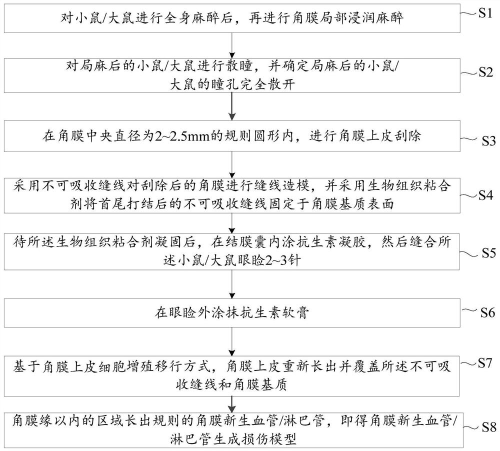
[0090] Operating procedures:

[0091] In step 1 (S1), after general anesthesia is performed on C57 adult mice, local corneal infiltration anesthesia is performed.

[0092] During specific implementation, a mixed solution of 3.3% tribromoethanol and tert-amyl alcohol (1 g: 1 ml) was prepared, and intraperitoneal anesthesia was performed at 0.024 ml / g body weight. C57 adult mice can be injected intraperitoneally with a 1ml syringe with a No. 4 needle. During intraperitoneal injection, hold the syringe in the right hand, grasp the tail of the C57 adult mouse with the little finger and ring finger of the left hand, and grasp the neck of the C57 adult mouse with the other three fingers, so that the head of the C57 adult mouse is down. In this way, the organs in...

Embodiment 2

[0117] The construction method of this embodiment is similar to the construction method of embodiment 1, and the differences are as follows:

[0118] In step 1, after 3 min of intraperitoneal injection of anesthesia, complete animal general anesthesia.

[0119] In step 2, the pupil dilation time is 3 minutes, and after 3 minutes, the pupils can be completely dilated under the light under a stereomicroscope.

[0120]In step 3, when the cornea is scraped mechanically, it is scraped to the superficial stromal layer; the specific mechanical scraping operation is: use alcohol to assist and use a 2mm marker ring to place an indentation in the center of the cornea, inject 30ul of 20% ethanol into the ring, and after 30s Absorb the remaining ethanol with a dry blood-sucking sponge, immediately rinse the corneal epithelium with balanced solution or saline, and mechanically scrape off the regular circular corneal epithelium with a diameter of 2.5 mm to expose the superficial stroma.

...

Embodiment 3

[0127] The construction method of this embodiment is similar to the construction method of embodiment 1, and the differences are as follows:

[0128] SD adult rats were selected in this embodiment, and a rat corneal neovascularization / lymphangiogenesis injury model was constructed.

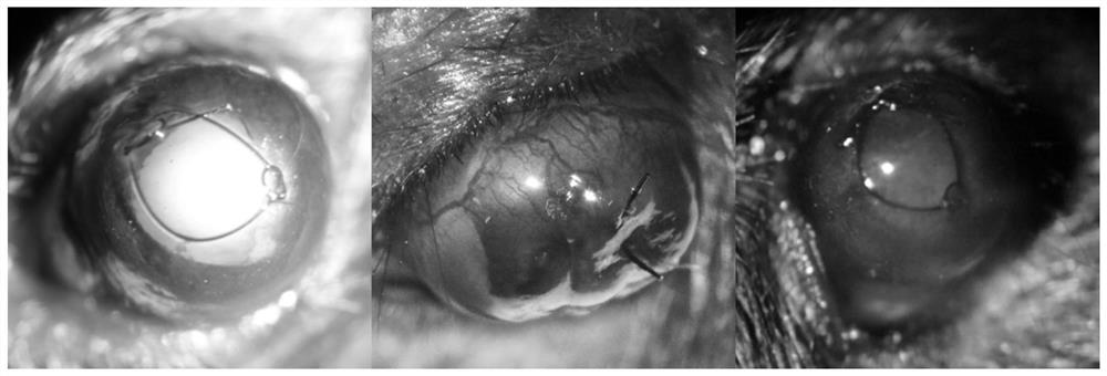
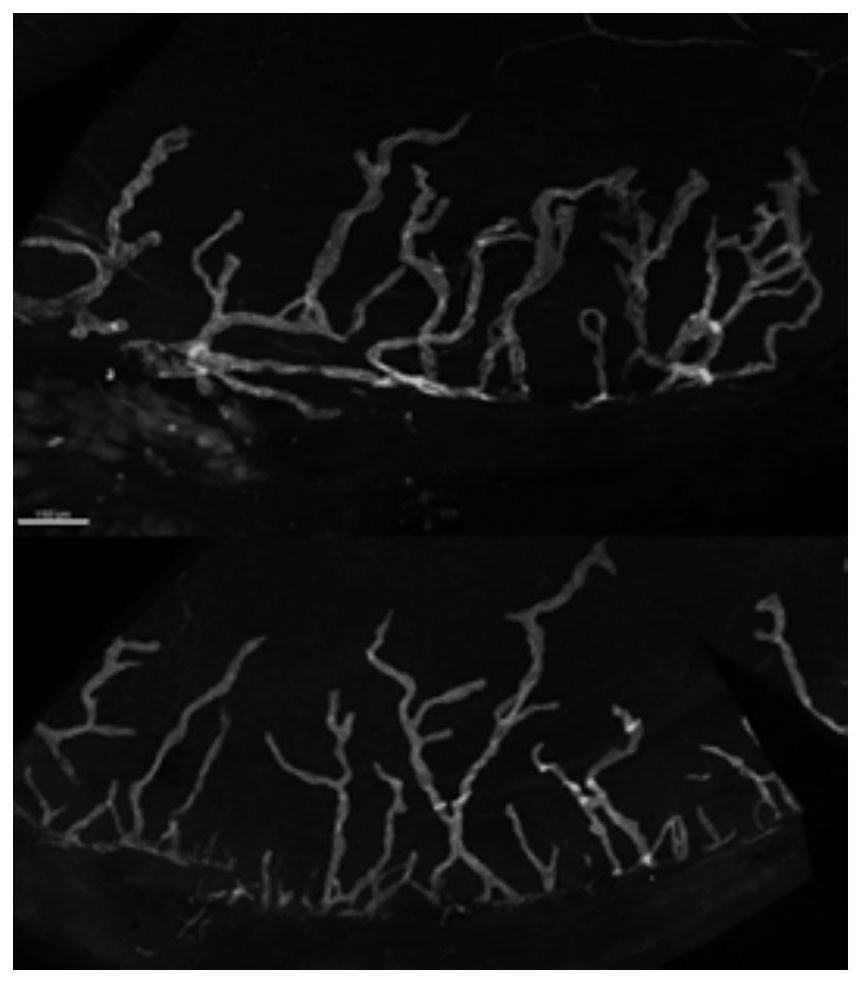
[0129] The rat corneal neovascularization / lymphangiogenesis injury model constructed in this example, its new blood vessel map under the slit lamp 14 days after the suture operation, the lymphatic vessel (LYVE1) immunofluorescence staining map and the model 14 days after the suture operation The diagrams in the damage process are the same as those shown in Example 1, and will not be repeated in this example.


PUM


Abstract

The invention provides a corneal neovascularization / lymphatic vessel generation injury model and a construction method and application thereof. The model is constructed on the basis of a corneal suture thought by combining mechanical scraping of corneal epithelium and bonding of the corneal suture to the corneal stroma by biological glue, so that the problems of high modeling difficulty and low efficiency when the corneal suture model is applied to small rodents are solved; and common reasons such as cornea perforation and suture shedding which cause modeling failure are avoided, and the advantages of regular growth of cornea suture neovascularization, avoidance of chemical reagent influence in a chemical induction method, single and controllable injury mode and the like are reserved. Besides, based on the construction method of the model, the model can be simply, effectively and controllably used as a novel effective model for mouse and rat corneal neovascularization or lymphatic vessel generation, so that the model is relatively high in applicability.

Description

technical field [0001] The invention relates to the field of organic compound synthesis, and mainly relates to a corneal neovascularization / lymphangiogenesis injury model and its construction method and application. Background technique [0002] Angiogenesis is a natural physiological process, and according to the location and time of occurrence, angiogenesis has different effects on the body. [0003] In most tissues of the human body, it is involved in the growth of tissue cells and the repair of damaged tissues. Under physiological conditions, the capillary network only grows around the limbus and does not extend to the cornea, and the cornea remains transparent. Under adverse conditions such as inflammation, trauma, and surgery, capillaries will invade the cornea, resulting in pathological CNV (ie, pathological corneal neovascularization). Its pathological process is mainly related to cytokines acting on blood vessels: cytokines acting on blood vessels play a key regul...

Claims


Application Information

Patent Timeline
Patent Type & Authority Applications(China)
IPC IPC(8): A61D1/00A61K49/00
CPCA61K49/0008A61D1/00
Inventor 李宗源王丽强黄一飞
Owner GENERAL HOSPITAL OF PLA
Why Eureka
Try Eureka