Preparation and application of fluorescence immunochromatography test strip for exosome quantification

A technology of fluorescent immunochromatography and test strips, which is applied in the direction of measuring devices, analytical materials, instruments, etc., can solve the problems of inconvenient downstream experiments, inability to remove pollution, expensive instruments, etc., and achieve simple, convenient and accurate exosomes in the experimental process Concentration, simple effect of equipment

Pending Publication Date: 2021-10-29
瑞太生物科技(沈阳)有限公司
View PDF4 Cites 1 Cited by
  • Summary
  • Abstract
  • Description
  • Claims
  • Application Information

AI Technical Summary

Problems solved by technology

[0003] The detection and quantification of exosomes is a routine experiment in exosome research. At present, there are many detection and quantification techniques that can be used for exosomes, such as dynamic light scattering technology, nanoparticle size tracer analysis technology, and adjustable resistance pulse transmission. Sensitive technology, flow cytometry and nano flow technology are often used to measure the concentration of exosomes, but these technologies also have certain limitations, among which dynamic light scattering technology, nanoparticle size tracer analysis technology, Adjustable resistance pulse sensing technology cannot remove the pollution of non-exosome particles in complex samples, resulting in unrealistic exosome concentration data. The detection sensitivity of flow cytometry is about 500nm, which cannot directly detect exosomes. Immunomagnetic beads can only be used as carriers to perform immunocapture of exosomes, and then carry out concentration detection. Because this process is too cumbersome, the operation is difficult, and the failure rate of the experiment is extremely high. Although nano flow technology can directly detect exosomes in samples The concentration of exosomes, but this instrument is too expensive, and ordinary laboratories do not have this instrument, resulting in the inability to quickly obtain concentration data for exosome samples to be tested, which brings inconvenience to downstream experiments

Method used


Image

  • Preparation and application of fluorescence immunochromatography test strip for exosome quantification
  • Preparation and application of fluorescence immunochromatography test strip for exosome quantification
  • Preparation and application of fluorescence immunochromatography test strip for exosome quantification

Examples


Embodiment 1

[0047] Example 1 A fluorescent immunochromatographic test strip for detecting exosomes provided by the present invention

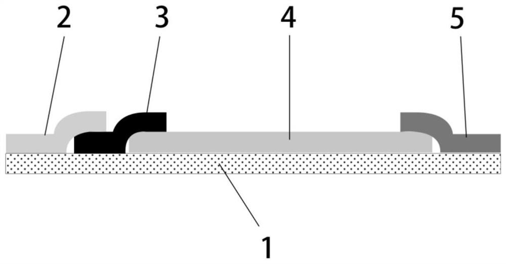
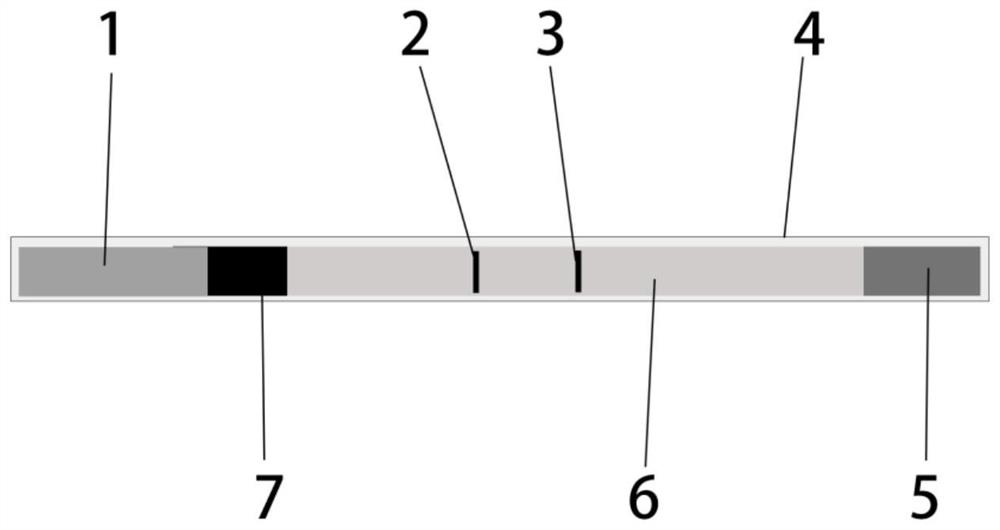
[0048] Such as figure 1 As shown, the fluorescent immunochromatographic test strip test for detecting exosomes includes a back plate and a sample pad pasted on the back plate, a binding pad, an NC film, and a water-absorbing pad, wherein the binding pad is connected to the sample pad and the NC film respectively. One end of the membrane is overlapped, and the other end of the NC membrane is overlapped with the absorbent pad. The NC membrane is a nitrocellulose membrane with a pore size of 450-700nm, and the sample pad and binding pad are polyester membranes.

[0049] Such as figure 2As shown, the NC membrane is sprayed with detection lines and quality control lines, and the binding pad is attached with fluorescent microsphere-labeled mouse anti-human CD9 antibody and fluorescent microsphere-labeled rabbit IgG. The fluorescent microspheres have a particl...

Embodiment 2

[0050] Example 2 The method of the fluorescent immunochromatographic test strip for detecting exosomes provided by the present invention

[0051] 1. CD9 Antibody and Rabbit IgG Fluorescent Microsphere Labeling

[0052] (1) Fluorescent microspheres are activated

[0053] Take 200ul of fluorescent microspheres with a solid content of 1% and add them to a centrifuge tube, and add 1ml of MES buffer solution (MES200mg / ml), mix well, put into a centrifuge, centrifuge at 6000g for 5min, and remove the supernatant. Then add 1ml of MES buffer (MES 200mg / ml) to the centrifuge tube to resuspend the fluorescent microspheres, then add 100ul (20mg / ml) EDC and 100ul (20mg / ml) NHS to the solution, shake and mix After 40 minutes, place the centrifuge tube in a centrifuge, centrifuge at 6000 g for 5 minutes, and suck off the supernatant.

[0054] (2) Fluorescent microsphere labeling of CD9 monoclonal antibody

[0055] Add 1ml of MES buffer (MES 200mg / ml) to the activated microspheres, mix by...

Embodiment 3

[0074] Example 3 The method provided by the present invention for detecting the concentration of exosomes using the fluorescent immunochromatographic test strip

[0075] Concentration detection of exosome samples

[0076] Step 1: Sample Preparation

[0077] Take an appropriate amount of exosome sample, add sample diluent to the exosome sample, and mix well to form an exosome sample mixture.

[0078] Step 2: Prepare test strips

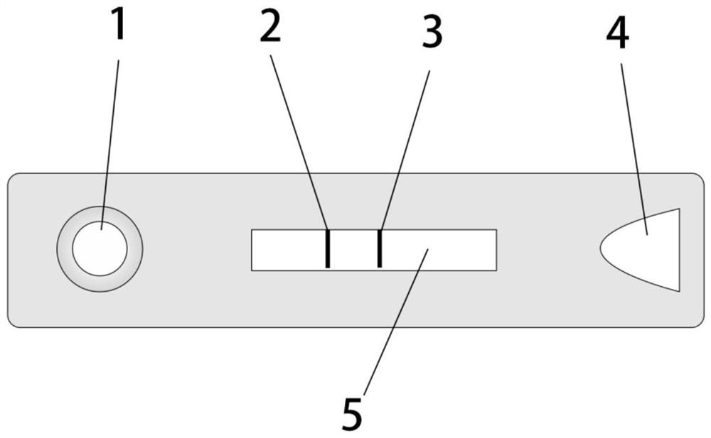
[0079] Remove the outer packaging of the test paper card sample, place the test paper card on a horizontal plane, and set it aside.

[0080] Step 3: Adding samples

[0081] Aspirate 80-100ul exosome sample mixture, add it to the sample slot of the test paper card, and time it.

[0082] Step 4: Data Detection

[0083] After 15 minutes, the test paper card was placed in a fluorescent immunoassay analyzer (purchased from Guangzhou Lanbo Biotechnology Co., Ltd.) to detect the concentration of exosomes.

[0084] Quantification of Exosome Concentration...


PUM

PropertyMeasurementUnit
pore sizeaaaaaaaaaa
particle sizeaaaaaaaaaa
diameteraaaaaaaaaa

Abstract

The invention discloses preparation and application of a fluorescence immunochromatography test strip for exosome quantification, and particularly relates to the field of biological detection. The test strip comprises a back plate, a sample pad, a combination pad, an NC membrane and a water absorption pad, wherein the sample pad, the combination pad, the NC membrane and the water absorption pad are pasted on the back plate, the combination pad is in lap joint with one end of the sample pad and one end of the NC membrane, the other end of the NC membrane is in lap joint with the water absorption pad, and a detection line and a quality control line are sprayed on the NC membrane. A mouse anti-human CD9 antibody marked by fluorescent microspheres and rabbit IgG marked by fluorescent microspheres are attached to the combination pad, a mouse anti-human CD9 monoclonal antibody is coated on the detection line, and a goat anti-rabbit antibody is coated on the quality control line. By utilizing the optimized fluorescence immunochromatography technology, the concentration of the exosome can be measured more conveniently, quickly and accurately, and a new technical means is provided for the detection of the concentration of the exosome.

Description

technical field [0001] The invention relates to the technical field of biological detection, in particular to the preparation and application of a fluorescent immunochromatographic test strip for quantifying exosomes. Background technique [0002] Exosomes are small vesicles with a phospholipid bilayer structure secreted by living cells, with a diameter of 30-150nm and a density of 1.13-1.19g / ml, which can exist in various body fluids, such as serum, plasma, saliva, urine fluid, ascites, spinal fluid, milk, etc. Exosomes contain a variety of biomolecules, such as mRNA, miRNA, proteins, lipids, etc., which can be delivered to recipient cells, thereby changing the physiological or pathological functions of recipient cells. In recent years, exosomes have attracted widespread attention as intercellular information transfer tools and biomarkers of various diseases. Exosomes have great potential in the fields of biomedicine and disease diagnosis. [0003] The detection and quant...

Claims


Application Information

Patent Timeline
Patent Type & Authority Applications(China)
IPC IPC(8): G01N33/569G01N33/558G01N33/543G01N33/533G01N33/577
CPCG01N33/56966G01N33/558G01N33/533G01N33/54313G01N33/577
Inventor 张海心
Owner 瑞太生物科技(沈阳)有限公司
Why Eureka
Try Eureka