Application of naringenin in preparation of accelerant for promoting polarization of M1 microglial cells to M2

A technology of microglia and naringenin, which is applied in the field of biomedicine to achieve the effects of removing toxic substances, inhibiting inflammatory reactions, and promoting transformation

Pending Publication Date: 2021-11-23
GUANGDONG OCEAN UNIVERSITY
View PDF2 Cites 0 Cited by
  • Summary
  • Abstract
  • Description
  • Claims
  • Application Information

AI Technical Summary

Problems solved by technology

Recent experiments have found that naringenin has a certain protective effect on cerebral ischemia-reperfusion injury and Aβ-induced neuron damage; however, there is no report on the role of naringenin in the subtype differentiation of microglial cells

Method used


Image

  • Application of naringenin in preparation of accelerant for promoting polarization of M1 microglial cells to M2
  • Application of naringenin in preparation of accelerant for promoting polarization of M1 microglial cells to M2
  • Application of naringenin in preparation of accelerant for promoting polarization of M1 microglial cells to M2

Examples


Embodiment 1

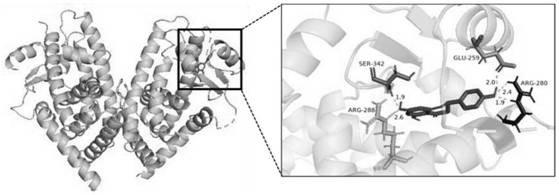
[0028] Example 1 Molecular docking study of naringenin and its target PPARγ

[0029] Method: The 3D structure of the compound naringenin was obtained from PubChem (https: / / pubchem.ncbi.nlm.nih.gov / ), and the core was screened from the RCSB PDB (http: / / www.rcsb.org / ) database For the crystal structure of the target protein, use pymol (version 2.4.0) to remove water molecules and small ligand molecules in the protein, and isolate the protein structure. The genetic algorithm in AutoDock 4.2.6 software was used for semi-flexible docking. The coordinates and size of the center of the box were set according to the position of the active site of the protein molecule and the area where it may act on the small molecule of the ligand. The rest of the parameters were kept at default. For the compound Molecular docking analysis with the target protein.

[0030] Results: Molecular docking is to simulate the interaction between the ligand small molecule and the receptor biomacromolecule, o...

Embodiment 2

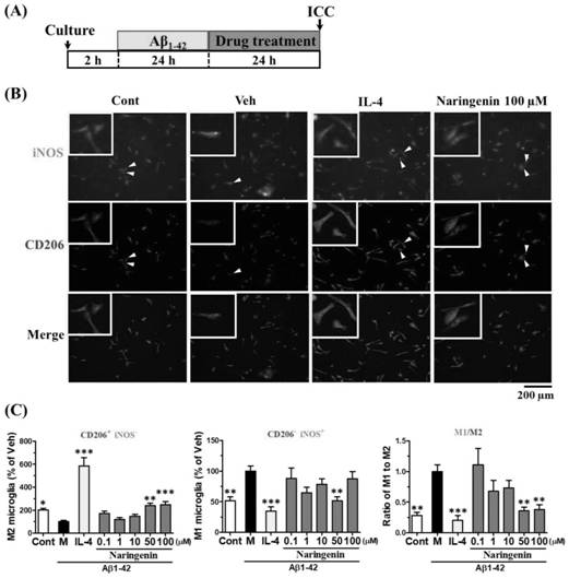
[0031] Example 2 Naringenin promotes the polarization of M1 microglial cells to M2

[0032] Primary microglial cell culture: 5-6 ddY mice (P2-4), the cerebral cortex was separated after alcohol sterilization, the dura mater was removed, chopped and placed in DMEM (+FBS) medium, centrifuged (1000rpm, 3min), remove the supernatant, add 0.25% trypsin 2mL, at 37 degrees, 5% CO 2 Incubate in an incubator for 30min, add 4mL of 10% FBS-DMEM medium, centrifuge (2000rpm, 5min), remove the supernatant, add DNase I / Trypsine inhibitor 2mL, at 37 degrees, 10% CO 2 Incubate in an incubator for 15min, add 4mL of 10% FBS-DMEM medium, centrifuge (1000rpm, 3min), remove the supernatant, add 4mL of 10% FBS-DMEM medium, blow and dissociate the cells with a rubber dropper, Add 2 mL of 15% BSA in PBS solution from the bottom, centrifuge (2000 rpm, 6 min), remove the supernatant, add 2 mL of 10% FBS-DMEM medium, count the cells and inoculate them on a 6-cm culture dish (8*10 5 cells). After 14 da...

Embodiment 3

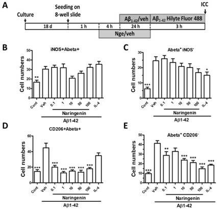
[0035] Example 3 Naringenin activates the phagocytosis of microglial cells on Aβ

[0036] Method: The experimental procedure is as follows image 3 As shown in A, the primary cultured microglial cells were seeded on 8-well plates (1.5*10 4 cells / well), add naringenin (0.1~100μM) or positive control IL4 after 1h, add Aβ1-42 (1μM) after 4 hours of culture and continue to culture for 24h, remove the supernatant containing Aβ and naringenin, add Aβ1 -42 Hilyte Fluor 488, cultured for 3 hours, fixed cells with 4% PFA, added primary antibody CD206 (1:200, M2 marker) or iNOS (1:50, M1 marker) and secondary antibody Alexa Fluor 594 (Invitrogen, Carlsbad, CA, USA) for immunofluorescence staining, photographed with a fluorescent microscope (BX-61 / DP70, Olympus) and analyzed with Metamorph software.

[0037] The result is as image 3 As shown in B, after the addition of Aβ1-42, the phagocytosis of fluorescently labeled Aβ1-42Hilyte Fluor488 by microglial cells was greatly increased, b...


PUM


Abstract

The invention discloses application of naringenin in preparation of an accelerant for promoting polarization of M1 microglial cells to M2. Researches show that the naringenin promotes polarization of A beta-induced M1 microglial cells to M2, and the addition of the naringenin activates the number of M2 microglial cells, so that A beta degrading enzyme is highly expressed; and further researches show that the naringenin participates in the transformation of M2 microglial cell subtypes induced by the naringenin by up-regulating the expression of PPAR gamma. The naringenin has the effect of promoting the microglial cells to be transformed from M1 phenotype to M2 phenotype, and has a relatively great application prospect in the aspects of inhibiting inflammatory response, removing toxic substances, repairing injured nerves and the like.

Description

technical field [0001] The invention relates to the technical field of biomedicine, and more specifically relates to the application of naringenin in the preparation of an accelerator for promoting the polarization of M1 type microglial cells to M2. Background technique [0002] Microglia is the only cell derived from mesoderm in nervous tissue. Two types of activation of microglia are known. There are pro-inflammatory M1 phenotype microglia and anti-inflammatory M2 phenotype microglia. Pro-inflammatory M1 phenotype microglia produce interleukin-1β (IL-1β), tumor necrosis factor-α (TNF-α), inducible nitric oxide synthase (iNOS), etc. IL-1β is an effector. In the presence of IL-1β, microglia induce inflammation in the brain and release free radicals such as free radicals and reactive oxygen species. In addition, in IL-1β-producing microglia, the ability to engulf metabolic waste such as aged (destroyed) cells was reduced. Thus, pro-inflammatory M1 phenotype microglia dama...

Claims


Application Information

Patent Timeline
Patent Type & Authority Applications(China)
IPC IPC(8): A61K31/352A61P25/28A61P29/00
CPCA61K31/352A61P25/28A61P29/00
Inventor 杨志友张永平宋采邓嘉航丰心月马智慧
Owner GUANGDONG OCEAN UNIVERSITY
Why Eureka
Try Eureka