Pd-l1 fusion protein and use thereof

Inactive Publication Date: 2017-07-06
GENEXINE INC +1
View PDF0 Cites 14 Cited by
  • Summary
  • Abstract
  • Description
  • Claims
  • Application Information

AI Technical Summary

Benefits of technology

[0015]The fusion protein of the present invention, which comprises the extracellular domain of PD-L1 protein or a fragment thereof and a modified immunoglobulin Fc region, has characteristics in that it has higher stability than conventional Ig fusion proteins and can be produced in large amounts. In addition, it can inhibit cytokine production and cell proliferation of T cells, and has the effects of controlling regulatory T cells (Treg) that can inhibit the function of abnormally activated immune cells and control inflammatory responses.
[0016]Therefore, the fusion protein of the present invention can be effectively use

Problems solved by technology

However, a T cell immune tolerance-based immunotherapeutic agent using an agonist has not yet been developed.
However, IgG1 used in conventional Ig fusion technologies cause antibody dependent cell-mediated cytotoxicity (ADCC) and complement dependent cytotoxicity (CDC

Method used


Image

  • Pd-l1 fusion protein and use thereof
  • Pd-l1 fusion protein and use thereof
  • Pd-l1 fusion protein and use thereof

Examples


Example

Example 1: Preparati
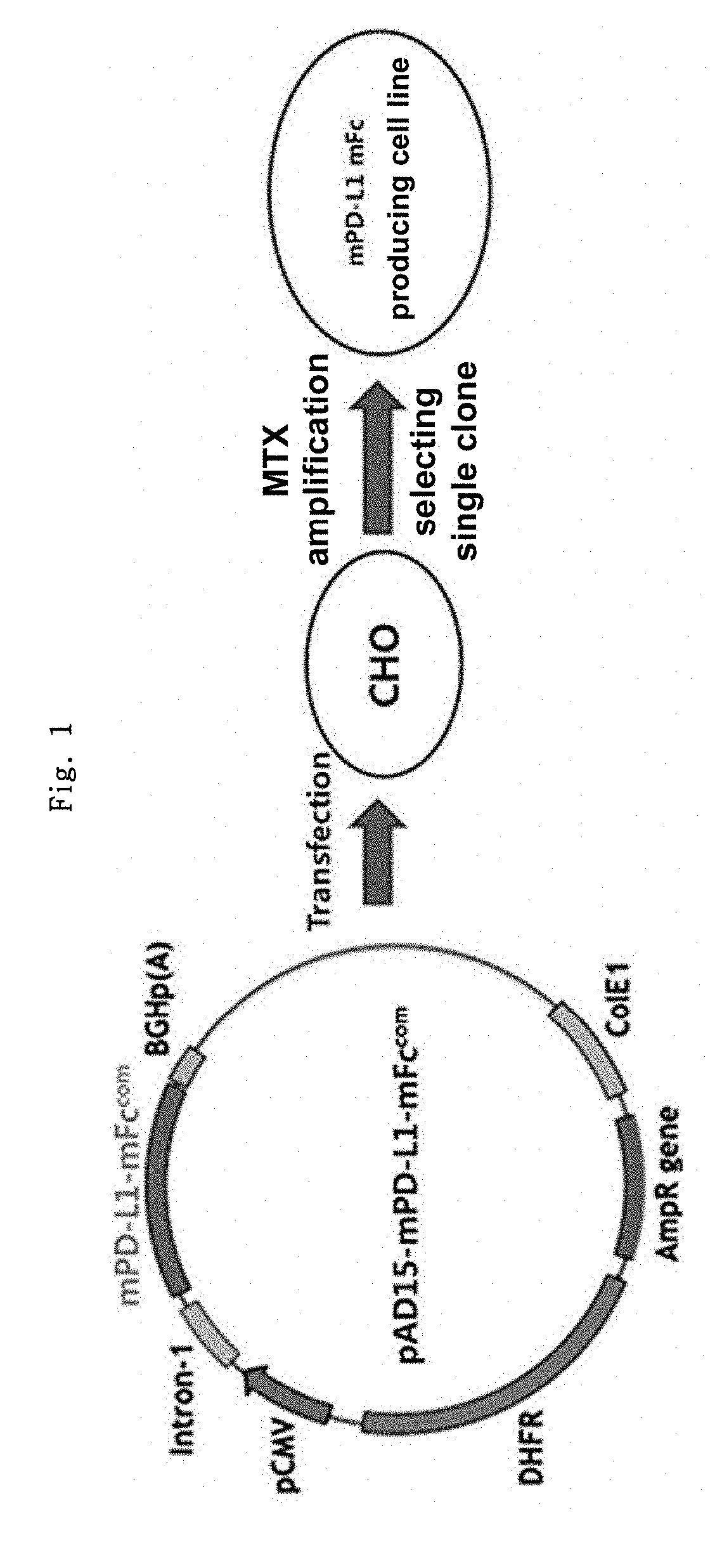
on of mPD-L1-mFc (Mouse PD-L1-Mouse Non-Lytic IgG2a) Gene Construct

[0155]Mouse PD-L1 protein (mPD-L1) and human PD-L1 protein (hPD-L1) have a sequence homology of about 70%. Thus, when human PD-L1 protein is repeatedly administered to a mouse, an antibody (anti-drug antibody, ADA) against the human PD-L1 protein might be developed, which could make it difficult to predict the accurate efficacy of the human PD-L1 protein, in some cases.

[0156]Thus, before an experiment was conducted using a human PD-L1-Fc fusion protein, the effect of a mouse PD-L1-Fc fusion protein was analyzed by an in vivo experiment in mice. Non-lytic Fc (hereinafter referred to as “mFc”) was constructed to provide a gene construct capable of producing mPD-L1-mFc, and a cell line expressing mPD-L1-mFc was constructed. The recombinant protein-expressing cell line was suspension-cultured, and the culture medium was collected, after which the recombinant protein was recovered by column purificatio...

Example

Example 2: Production of mPD-L1-mFc Protein

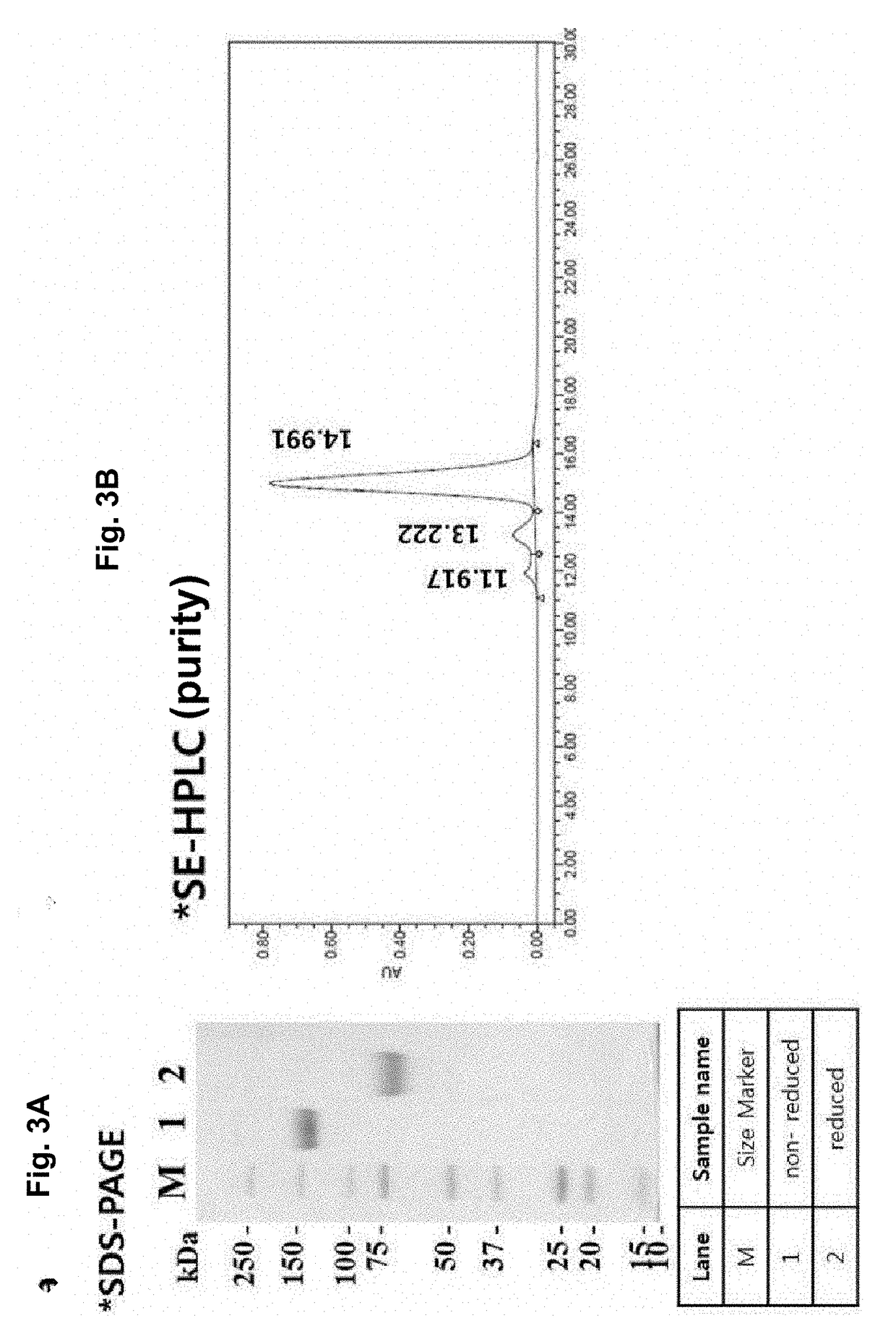
[0159]To produce the mPD-L1-mFc (mouse PD-L1-mouse non-lytic IgG2a) protein (SEQ ID NO: 10) in large amounts, the target protein was separated and purified from the cell culture medium produced by the mPD-L1-mFc suspension cell line obtained in Example 1.

[0160]To purify the protein, the protein purification process was monitored with time at a UV wavelength of 280 nm. As a result, elution of the target protein was verified by the peak that appeared when elution buffer (0.1M glycine, pH 3.0) passed through the protein A resin column (FIG. 3). In addition, the purified mPD-L1-mFc protein was analyzed by SDS-PAGE, and as a result, the protein size was about 150 KDa in a non-reducing condition and about 75 KDa in a reducing condition, indicating that mPD-L1-mFc is in the form of a homodimer (FIG. 3a). In addition, the purification product was analyzed by SE-HPLC to determine its purity (FIG. 3b), and the level of the impurity endotoxin was meas...

Example

Example 3: Evaluation of In Vitro Activity of mPD-L1-mFc Protein

[0161]To evaluate the activity of the mPD-L1-mFc protein (SEQ ID NO: 10) purified in Example 2, the immunosuppressive effect of the protein was analyzed in vitro using mouse splenocytes.

[0162]As schematically shown in FIG. 4, anti-CD3 and the mPD-L1-mFc protein were coated on microbeads at a ratio of 1:1 or 1:4. In addition, the beads and the mouse splenocytes were used at a ratio of 10:1 to stimulate the splenocytes (5×10⁶ beads: 5×10⁵ splenocytes).

[0163]Using the microbeads coated with the anti-CD3 antibody and the mPD-L1-mFc protein, the mouse splenocytes were stimulated in a microwell plate. After 48 hrs, the expression level of the cell proliferation factor Ki-67 in the mouse splenocytes was analyzed.

[0164]As a result, inhibition of the proliferation of the mouse splenocytes by the mPD-L1-mFc protein (reduction in Ki-67 expression) was observed, which indicates that the mPD-L1-mFc fusion protein has immunosuppressi...


PUM


Abstract

The present invention relates to a fusion protein composed of the extracellular domain of PD-L1 and a modified immunoglobulin Fc region. The extracellular domain of PD-L1 and a fragment thereof have excellent immunomodulatory activity, and can be used as an immunomodulatory agent if a modified immunoglobulin Fc region is coupled thereto. Accordingly, the PD-L1 fusion protein according to the present invention demonstrated its excellent effect in disease models of inflammatory bowel disease, colitis, psoriasis, asthma and arthritis, and thus can be very effectively used for the treatment of such diseases.

Description

TECHNICAL FIELD[0001]The present invention relates to a PD-L1 fusion protein prepared by coupling PD-L1 to an immunoglobulin Fc region, which has increased stability and activity. Moreover, the present invention relates to a pharmaceutical composition comprising PD-L1 or a specific fragment thereof, and more particularly, to a pharmaceutical composition for treating an immune disease, which comprises PD-L1 or a specific fragment thereof.BACKGROUND ART[0002]Human hPD-L1 (human Programmed Cell Death-Ligand 1), a ligand for PD-1 (programmed death-1), is a type 1 transmembrane protein expressed not only in hematopoietic cells such as T lymphocytes, B lymphocytes, dendritic cells, or macrophages, but also in non-hematopoietic cells such as keratinocytes, islet cells, or hepatocytes.[0003]It is known that PD-1 binding to PD-L1 is expressed mainly in activated T cells and B cells, macrophages, or dendritic cells, and binds to PD-L1 to inhibit the cytokine production and cell proliferation ...

Claims


Application Information

Patent Timeline
IPC IPC(8): A61K38/17A61K39/00
CPCA61K38/17C07K2319/30A61K39/0005C07K14/70596A61K38/00C07K2317/52A61P1/04A61P17/06A61P19/02A61P29/00A61P37/02A61P37/06A61P3/10
Inventor SUNG, YOUNG CHULLEE, JI YEUNGSONG, MI-YOUNGLIM, HYE SEONGLEE, BYUNG HA
Owner GENEXINE INC
Why Eureka
Try Eureka