Method for detecting genetic toxicity by using 3D hepatocyte in-vitro micronucleus cells

A genotoxicity and hepatocyte technology, applied in the field of genotoxicity detection, can solve problems such as poor correlation of human experiments, and achieve the effect of ensuring accuracy and validity

Active Publication Date: 2021-12-10
SHANGHAI INNOSTAR BIO TECH
View PDF4 Cites 1 Cited by
  • Summary
  • Abstract
  • Description
  • Claims
  • Application Information

AI Technical Summary

Problems solved by technology

[0011] The technical problem to be solved in the present invention is to provide a method for detecting genotoxicity using 3D hepatic micronucleus cytomics in vitro in view of the shortcomings of the prior art for detecting genotoxicity in vitro and the human body test

Method used


Image

  • Method for detecting genetic toxicity by using 3D hepatocyte in-vitro micronucleus cells
  • Method for detecting genetic toxicity by using 3D hepatocyte in-vitro micronucleus cells
  • Method for detecting genetic toxicity by using 3D hepatocyte in-vitro micronucleus cells

Examples


Embodiment 1

[0059] Example 1 Cell Culture and Compound Treatment

[0060] The determination of CBMN-cyt was carried out according to the method recommended in the guidelines of in vitro mammalian cell micronucleus assay (OECD TG487, 2016) (FENECH M. Nature Protocols, 2007, 2(5): 1084-1104).

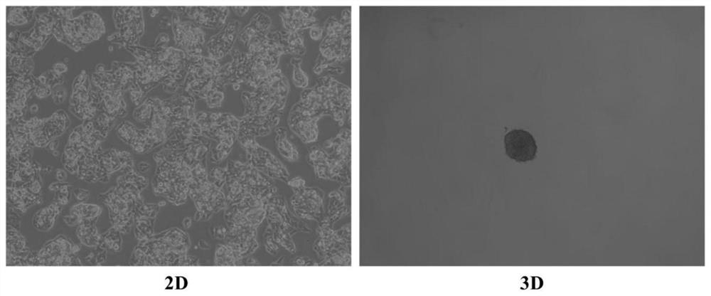
[0061] 1.1 2D cell culture and treatment

[0062] HepG2 cells in the logarithmic growth phase were trypsinized to make a single cell suspension and 1×10 5 The density of cells / well was seeded in 6-well cell culture plate, 37℃±1℃, 5±1%CO 2 Humidified cultivation. After 24 hours, the original medium was sucked off, and the prepared culture solution containing the compound and cyto-B at a final concentration of 5 μg / mL was added. For each treatment group and each condition, 2 wells were treated in parallel, and the sample volume was 4 mL. At the same time, DMEM medium containing vehicle and a final concentration of 5 μg / mL cyto-B was set as the control group. The culture solution was discarded afte...

Embodiment 2

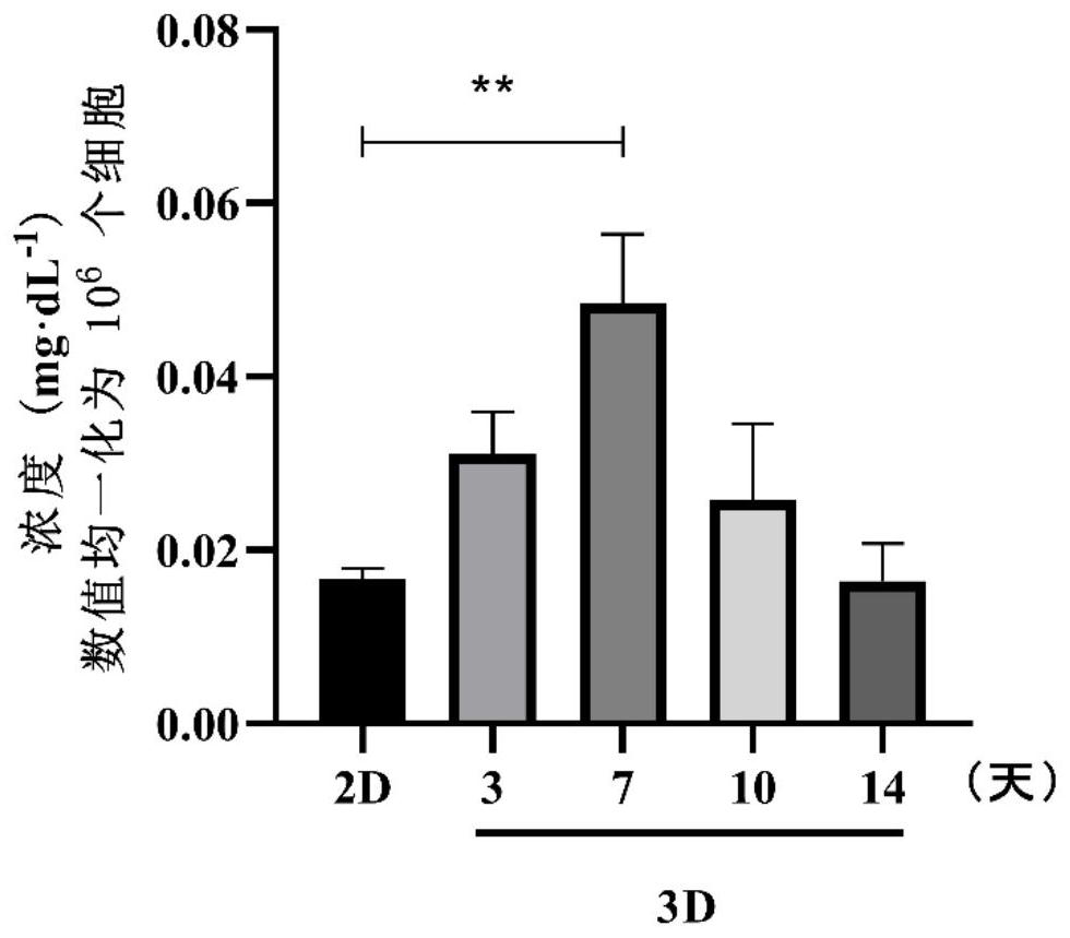
[0120] Example 2 Verification of 3D hepatocyte micronucleus cytomics test method

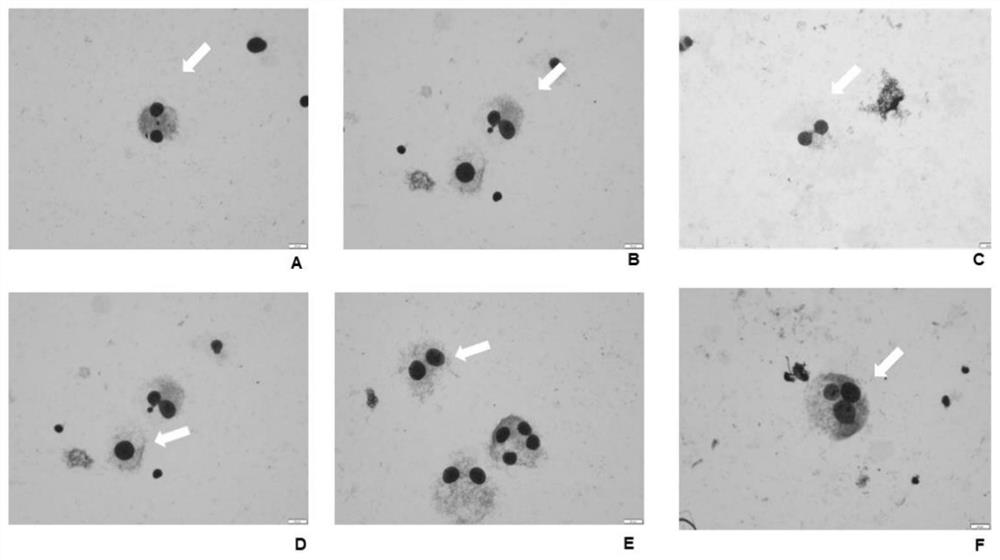
[0121] The present invention selects two genotoxic compounds benzopyrene (BaP) and cyclophosphamide (CPA) that require metabolic activation according to the recommendation of the guidelines for in vitro mammalian cell micronucleus test (OECD TG487, 2016), an aneuploid The inducer colchicine (COL), two suspected genotoxic compounds (-)-epigallocatechin gallate (EGCG) and furfuryl thioacetate, and a non-genotoxic compound amiodarone, had negative effects on established The 3D hepatocyte micronucleus cytomics assay method was validated to explore the applicability of this method for compounds with different mechanisms.

[0122] In this example, 6 representative substances with different genotoxicity mechanisms were selected to conduct a preliminary verification of the in vitro 3D hepatocyte micronucleus cytomics method, and the experimental results were compared with the micronucleus cytomics resul...


PUM


Abstract

The invention discloses a method for detecting genetic toxicity by using 3D hepatocyte in-vitro micronucleus cells. The method comprises the following steps of (1) constructing a 3D hepatocyte model in vitro by using common humanized hepatocytes HepG2 as a cell material by adopting a scaffold-free culture method combining hanging drops and ultra-low adsorption culture; and (2) carrying out micronucleus cell detection on a compound by using the 3D hepatocyte model, and analyzing the genetic toxicity mechanism of the compound. The in-vitro detection method can be used for analyzing various genetic toxicity mechanisms of the compound, improving the capability of predicting the genetic toxicity of drugs by an in-vitro cell model and providing information related to human body tests.

Description

technical field [0001] The invention belongs to the field of genotoxicity detection, and in particular relates to a method for genotoxicity detection by micronucleus cytomics in vitro of 3D liver cells. Background technique [0002] The current 3D cell culture technology has great development potential in toxicology research. Due to the increasing emphasis on animal ethics in the scientific research field, the use of 3D cell culture technology meets the requirements of the 3R principle of "reduce, replace, optimize". And because there are species differences between experimental animals (such as rodents) commonly used in drug toxicity evaluation and humans, the use of humanized cell models for drug toxicity evaluation can provide more human-related reference data in the early screening stage of drugs, shortening the development time and reduce R&D costs. 3D cell models currently used in genotoxicity tests include 3D skin models, 3D liver models, 3D airway models, etc. Sin...

Claims


Application Information

Patent Timeline
Patent Type & Authority Applications(China)
IPC IPC(8): C12Q1/02C12N5/071
CPCG01N33/5014G01N33/5067C12N5/067C12N2509/00C12N2513/00
Inventor 王敬亭赵泽浩赵田田徐静怡李英奇周长慧常艳
Owner SHANGHAI INNOSTAR BIO TECH
Why Eureka
Try Eureka