Chemical embryotoxicity prediction model and establishment method thereof

A technology of embryotoxicity and prediction model, which is applied in the field of prediction of embryotoxicity of new chemicals, and can solve the problems of many influencing factors, long research and experiment cycles, etc.

Pending Publication Date: 2022-01-14
SHANGHAI JIAOTONG UNIV SCHOOL OF MEDICINE
View PDF0 Cites 0 Cited by
  • Summary
  • Abstract
  • Description
  • Claims
  • Application Information

AI Technical Summary

Problems solved by technology

However, there are several problems that cannot be ignored in carrying out animal experiments: 1. It requires a lot of financial resources; 2. The experiment cycle is long; 3. It requires a large number of experimental animals; 4. It is difficult to carry out metabolism and mechanism research due to many influencing factors in the body
However, currently existing ESTs only use the gene Myh6, which encodes the heavy chain of myocardin, as the molecular endpoint. Whether the expression of other genes can also be used as molecular endpoints for EST to predict the embryotoxicity of chemicals remains to be confirmed.

Method used


Image

  • Chemical embryotoxicity prediction model and establishment method thereof
  • Chemical embryotoxicity prediction model and establishment method thereof
  • Chemical embryotoxicity prediction model and establishment method thereof

Examples


Embodiment 1

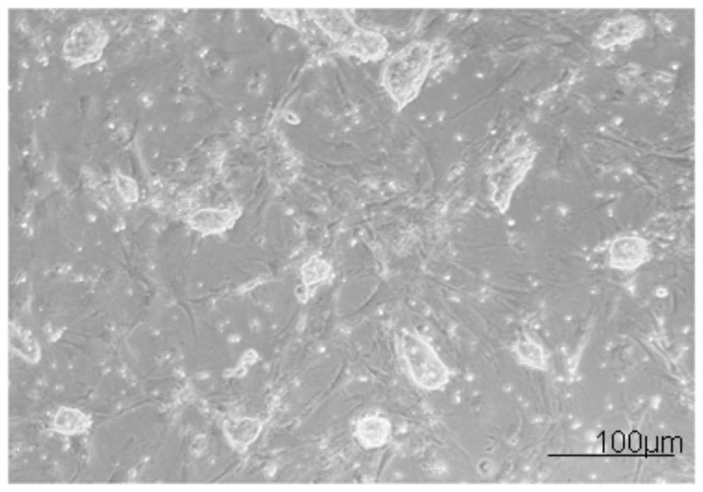
[0099] Example 1: Isolation and culture of mouse embryonic fibroblasts

[0100] In the present invention, mouse embryonic fibroblasts (MEF) are used as trophoblast cells for culturing ES.

[0101] 1.1 Isolation method of MEF primary cells:

[0102] (1) Take a beaker filled with 50ml of 75% ethanol, soak scissors, tweezers and other surgical instruments, the soaked instruments are roasted and sterilized by an alcohol lamp, then cooled and set aside.

[0103] (2) Prepare several 60mm-diameter bacterial culture dishes, which are respectively numbered as No. 1 dish, No. 2 dish, and No. 3 dish, and put into appropriate amount of high-temperature and high-pressure sterilized PBS, and add appropriate amount of penicillin-streptomycin to them solution. Among them, dish No. 1 contained the uterus taken out during the dissection of pregnant mice, dish No. 2 contained embryos with torn membranes, and dish No. 3 contained embryo trunks after treatment.

[0104] (3) After the pregnant m...

Embodiment 2

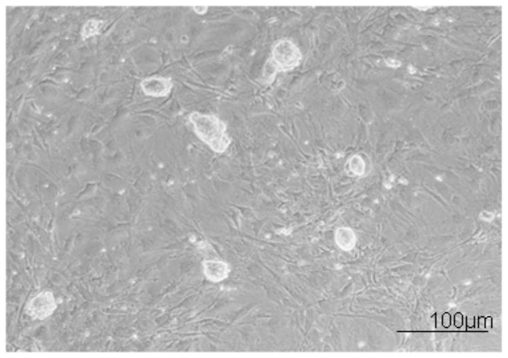
[0116] Example 2: In vitro expansion and culture of mouse ES

[0117] 2.1 Preparation of trophoblast cells

[0118] (1) Select MEF cells with appropriate algebra and density, add mitomycin C to the cell culture medium at a working concentration of 10 μg / ml, mix well and place in an incubator to continue culturing.

[0119] (2) After 2 hours, the culture medium containing mitomycin C was aspirated, and washed with PBS sterilized by high temperature and high pressure for 5 times, 5 minutes each time.

[0120] (3) After washing, replace with fresh culture medium to recover ES cells or continue culturing. The treated trophoblast cells supported the growth and maintenance of the undifferentiated state of mouse ES cells for up to 5 days.

[0121] 2.2 Recovery of ES cells

[0122] (1) Take out the ES cell cryopreservation tube from the liquid nitrogen, put it in a 37°C water bath to dissolve, take it out when it dissolves to the state of small ice crystals, and put it into the ult...

Embodiment 3

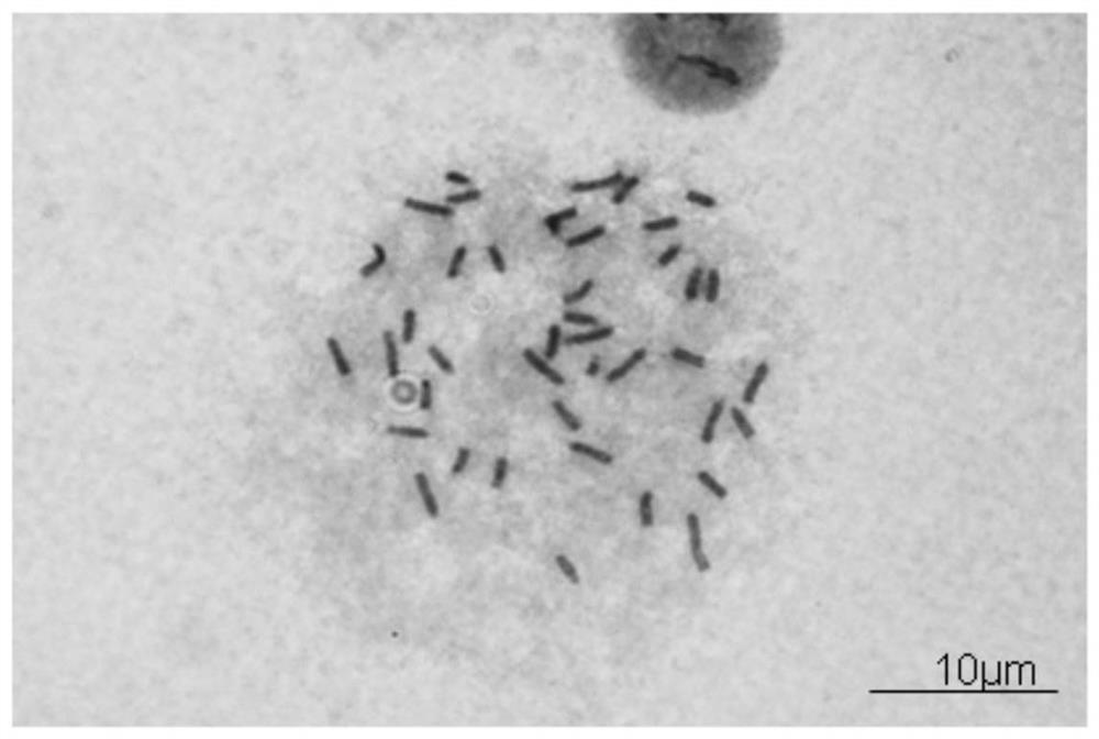
[0133] Example 3: Karyotype analysis and gender identification of mouse ES

[0134] 3.1 Karyotype analysis

[0135] (1) ES cells are cultured on a gelatin-coated petri dish, and the follow-up operations are carried out after the cells cover more than 80% of the petri dish;

[0136] (2) According to the results of the growth curve described in 2.4, add 50 ng / ml colchicine in the logarithmic growth phase, and continue culturing for 2 hours.

[0137] (3) Add EDTA-trypsin digestion solution after sucking up the culture solution, place it in a constant temperature incubator at 37°C for 5 minutes, blow gently with a pipette, then collect the cell suspension into a graduated centrifuge tube, and store at room temperature Centrifuge at 800 rpm for 5 minutes.

[0138] (4) Use a pipette to remove the supernatant, leaving about 0.1ml, then add hypotonic solution 0.56% w / v KCl to make up to 1ml, and put it in a water bath for 5 minutes.

[0139] (5) Add methanol: glacial acetic acid = ...


PUM

PropertyMeasurementUnit
diameteraaaaaaaaaa

Abstract

The chemical embryotoxicity prediction model comprises a cell type, wherein the cell type includes myocardial cells induced and differentiated by SP3 ES cells of which the karyotype is XX. The establishment method of the chemical embryotoxicity prediction model comprises the following steps of 1, separating and culturing mouse embryo fibroblasts; 2, carrying out in-vitro amplification culture on the mouse ES; 3, carrying out karyotype analysis and sex identification on the mouse ES; 4, maintaining and identifying the pluripotency of the mouse ES; 5, carrying out in-vitro induced differentiation on the mouse ES into myocardial cells; 6, performing immunochemical detection on the myocardial specific marker protein of the myocardial cells induced and differentiated by the mouse ES; 7, carrying out Real-time PCR detection of the expression profile of the myocardial cells induced and differentiated by the mouse ES; 8, selecting a mode compound; 9, detecting the cytotoxicity of the mode compound; 10, detecting the inhibition effect of the mode compound on the ES differentiation capability; and 11, evaluating the embryotoxicity of the model compound. The invention fills in a blank that no female mouse embryonic stem cells exist in an EST system.

Description

[0001] This application is a divisional application of the following applications: filing date: August 7, 2015; application number: 201510478375.3; invention title "a chemical embryotoxicity prediction model and its establishment method". technical field [0002] The present invention relates to a chemical embryotoxicity prediction model and its establishment method, in particular to a kind of mouse embryonic stem cell direction induction differentiation into cardiomyocytes with beating ability, and establishment of chemical embryotoxicity recognition on the obtained cardiomyocytes and prediction models, and applied to the prediction of embryotoxicity of new chemicals. Background technique [0003] At present, the toxicity data of chemicals are mostly obtained in experimental animals, such as acute toxicity test, subacute toxicity test, chronic toxicity test and so on. However, there are several problems that cannot be ignored in carrying out animal experiments: 1. It requir...

Claims


Application Information

Patent Timeline
Patent Type & Authority Applications(China)
IPC IPC(8): C12N5/077C12N5/0735C12N5/071C12Q1/02C12Q1/6851
Inventor 王艳程薇胡庆亮张星
Owner SHANGHAI JIAOTONG UNIV SCHOOL OF MEDICINE
Why Eureka
Try Eureka