Image processing method based on X-ray energy spectrum micro-area surface scanning

An image processing and X-ray technology, applied in the field of element distribution characterization involving scanning electron microscope-X-ray energy spectrometer, can solve the problems of inability to selectively enlarge the difference, the effective information is not prominent, the image is not clear, etc., to achieve accurate information enhancement High degree of accuracy, accurate results, and enhanced image recognition effect

Pending Publication Date: 2022-03-18
AVIC BEIJING INST OF AERONAUTICAL MATERIALS
View PDF0 Cites 1 Cited by
  • Summary
  • Abstract
  • Description
  • Claims
  • Application Information

AI Technical Summary

Problems solved by technology

[0003] At present, when users use scanning electron microscopes and X-ray energy spectrometers to perform surface scanning to obtain the surface distribution of elements, the common problem is that the signal-to-noise ratio is low, resulting in unclear images and inconspicuous effective information.
[0004] At present, most of the follow-up image enhancement method

Method used


Image

  • Image processing method based on X-ray energy spectrum micro-area surface scanning
  • Image processing method based on X-ray energy spectrum micro-area surface scanning
  • Image processing method based on X-ray energy spectrum micro-area surface scanning

Examples


Embodiment 1

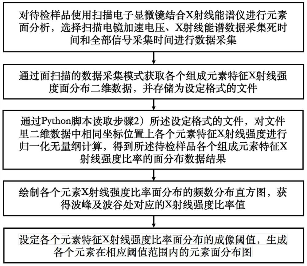
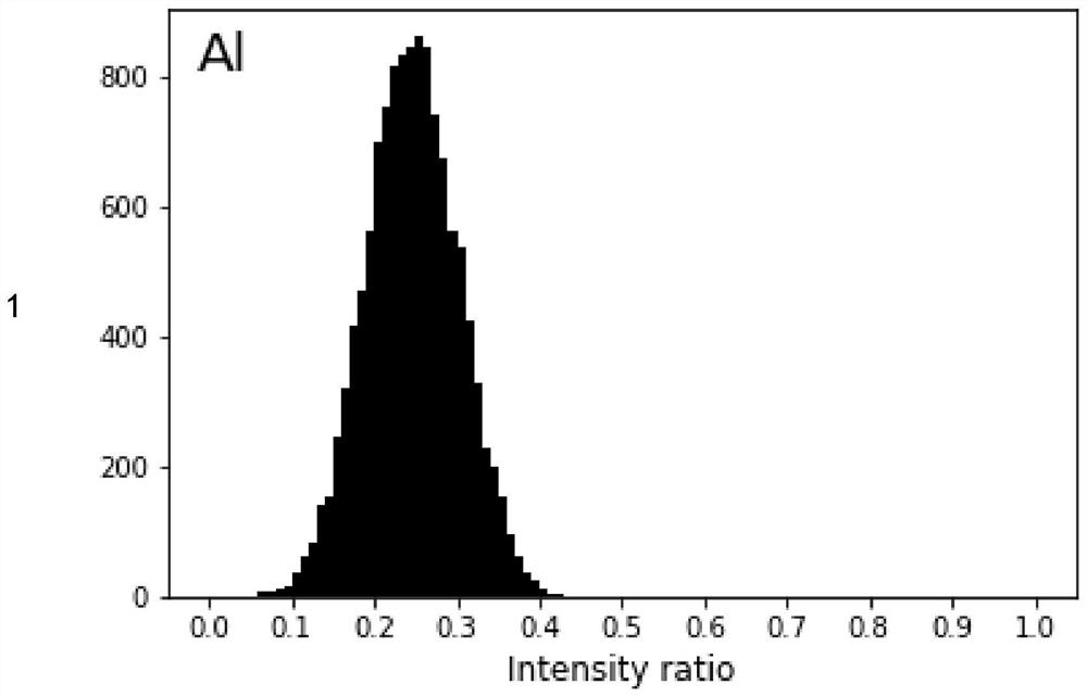
[0082] This embodiment provides an image processing method applied to an alloy sample containing three elements of aluminum, magnesium, and zinc based on X-ray energy spectrum micro-area scanning, but is not limited thereto, including the following steps:

[0083] Step 1) Use a scanning electron microscope in combination with an X-ray energy spectrometer to analyze the element surface of the sample to be inspected, and select the scanning electron microscope acceleration voltage, the X-ray energy spectrum data acquisition dead time and the entire signal acquisition time for data acquisition; where the sample to be inspected is Samples with good electrical conductivity that can be detected by a scanning electron microscope combined with an X-ray energy spectrometer; the energy of the incident electron beam of the scanning electron microscope used in the experiment is not lower than 2 times the energy value of the highest characteristic X-ray in the sample to be tested ; The dead...

Embodiment 2

[0101] This embodiment provides an image processing method applied to an alloy sample containing four elements of aluminum, copper, magnesium, and zinc based on X-ray energy spectrum micro-area scanning, but is not limited thereto, including the following steps:

[0102] Step 1) Use a scanning electron microscope in combination with an X-ray energy spectrometer to analyze the element surface of the sample to be inspected, and select the scanning electron microscope acceleration voltage, the X-ray energy spectrum data acquisition dead time and the entire signal acquisition time for data acquisition; where the sample to be inspected is Samples with good electrical conductivity that can be detected by a scanning electron microscope combined with an X-ray energy spectrometer; the energy of the incident electron beam of the scanning electron microscope used in the experiment is not lower than 2 times the energy value of the highest characteristic X-ray in the sample to be tested ; T...

Embodiment 3

[0119] This embodiment provides an image processing method applied to nickel-based superalloy samples based on X-ray energy spectrum micro-area scanning, but is not limited thereto, including the following steps:

[0120] Step 1) Use a scanning electron microscope in combination with an X-ray energy spectrometer to analyze the element surface of the sample to be inspected, and select the scanning electron microscope acceleration voltage, the X-ray energy spectrum data acquisition dead time and the entire signal acquisition time for data acquisition; where the sample to be inspected is Samples with good electrical conductivity that can be detected by a scanning electron microscope combined with an X-ray energy spectrometer; the energy of the incident electron beam of the scanning electron microscope used in the experiment is not lower than 2 times the energy value of the highest characteristic X-ray in the sample to be tested ; The dead time of the X-ray energy spectrometer is i...


PUM


Abstract

The invention provides an image processing method based on X-ray energy spectrum micro-area surface scanning, and belongs to the field of scanning electron microscope-X-ray energy spectrometer element distribution characterization. According to the method, intensity normalization calculation is carried out on element distribution data of X-ray energy spectrum surface scanning to obtain a distribution intensity ratio frequency distribution histogram of each element, and an imaging threshold value is set, so that the problems of low signal-to-noise ratio, unclear image, high resolution and the like when a scanning electron microscope and an X-ray energy spectrometer are used for micro-area surface scanning analysis in the prior art are solved. Effective information is not prominent, and the analysis efficiency is low.

Description

technical field [0001] The invention relates to the field of scanning electron microscope-X-ray energy spectrometer element distribution characterization, in particular to an image processing method based on X-ray energy spectrum micro-area scanning. Background technique [0002] Scanning Electron Microscope (SEM) equipped with X-ray Spectroscopy (Energy Dispersive X-Ray Spectroscopy; EDS) is one of the important characterization means for elemental analysis of material micro-regions. Scanning electron microscope has many advantages such as high image resolution (3nm~0.6nm), large depth of field (10 times that of optical electron microscope), non-destructive analysis and simple sample preparation, etc. The sample can be natural surface, fracture, block, reflective And translucent light film, etc. Equipped with X-ray energy spectrometer, it can comprehensively analyze the surface morphology and chemical composition and other information. X-ray energy spectrometer has three ...

Claims


Application Information

Patent Timeline
IPC IPC(8): G01N23/2273
CPCG01N23/2273
Inventor 滕超逸郑真刘昌奎
Owner AVIC BEIJING INST OF AERONAUTICAL MATERIALS
Why Eureka
Try Eureka