Purification process and application of antigen capable of being used in diagnosis of multiple myitis/dermatomyositis

A purification method and dermatomyositis technology, applied to the antigen purification process and application field that can be used for the diagnosis of polymyositis/dermatomyositis, can solve the problems of long time consumption, high cost, low yield, etc.

Inactive Publication Date: 2006-03-01
SINOGENOMAX +1
View PDF0 Cites 3 Cited by
  • Summary
  • Abstract
  • Description
  • Claims
  • Application Information

AI Technical Summary

Problems solved by technology

[0009] Over the past 20 years, many authors have made a lot of efforts on the purification of Jo-1 antigen, but the purification process is complicated, the yield is low, the cost is high, and it takes a long time. It is difficult to produce on a large scale to meet the needs of clinical laboratory diagnostic applications.
The successful purification of histidyl-tRNA synthetase by a small amount of affinity chromatography (see: Targoff IN and Reichlin M, J Immunol., 138:2874-82, 1987) was only found in laboratory applications and was not used for large-scale purification of Jo- 1 antigen

Method used


Image

  • Purification process and application of antigen capable of being used in diagnosis of multiple myitis/dermatomyositis
  • Purification process and application of antigen capable of being used in diagnosis of multiple myitis/dermatomyositis
  • Purification process and application of antigen capable of being used in diagnosis of multiple myitis/dermatomyositis

Examples


Embodiment 1

[0082] Extractable nuclear antigen extraction and manufacture of extractable nuclear antigen acetone powder

[0083] 1 Take fresh or -80°C frozen pig spleen and other animal tissues, and the frozen tissue needs to be thawed and recovered at 4°C overnight (more than 12 hours);

[0084] 2 Weigh 100g of porcine spleen and other tissues, add 200ml homogenization buffer (0.01M PBS, pH7.0, disodium edetate 0.01M, phenylmethylsulfonyl fluoride 1.5mM, dimercaptothreitol 1mM, Trypsin inhibitor 4μg / ml, sodium bisulfite 1.5mM), low-speed homogenization, 30 seconds / time, interval 2 minutes / time, ten times in total;

[0085] 3 Magnetically stir the tissue homogenate for 1-2 hours;

[0086] 4 Centrifuge at 4°C, 10,000G, for 1 hour;

[0087] 5 Discard the precipitate, take the supernatant and centrifuge at 4°C, 100,000G, for 1 hour;

[0088] 6 Discard the precipitate, put the supernatant in a beaker, and slowly add 5 times pre-cooled acetone while stirring;

[0089] 7 4°C, 10,000G, centr...

Embodiment 2

[0096] Fabrication of Immunoaffinity Columns

[0097] 1 Epoxy activation of agarose

[0098] 1) Rinse the dry agarose gel with deionized water (200ml / g) for 3 times, and drain it on the Buchner funnel;

[0099] 2) Transfer the washed gel into aqueous sodium hydroxide solution, then add epichlorohydrin, and their final concentrations in the activation system are respectively: gel 30% (v / v), epichlorohydrin 5% (v / v), sodium hydroxide 0.4M;

[0100] 3) shaking the above suspension under mild conditions, and reacting at 40°C for 2-3 hours;

[0101] 4) Transfer the activated gel into a Buchner funnel to drain, rinse the gel several times with deionized water, and set aside.

[0102] 2 Manufacture of Ligand

[0103] 1) Mix the monospecific Jo-1 antiserum with an equal amount of binding buffer (20 mM sodium phosphate buffer, pH=7.0), and filter with a 0.45 μm filter;

[0104] 2) Load the above-mentioned treated antiserum onto the rProtein A affinity column, and collect the loadi...

Embodiment 3

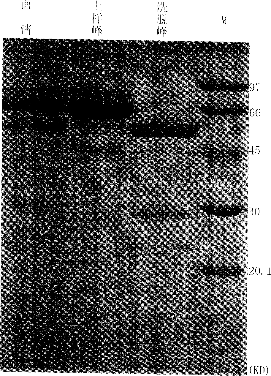
[0126] Purification of Jo-1 Antigen by Immunoaffinity Chromatography

[0127] 1) The extractable nuclear antigen solution is dialyzed against the binding buffer for 16-24 hours, and the solution is changed at least 5 times during the dialysis process. If it is an extractable nuclear antigen acetone powder, first use the extraction buffer to extract the extractable content of the acetone powder. nuclear antigen, redialysis;

[0128] 2) Filter the extractable nuclear antigen solution with a 0.45 μm filter,

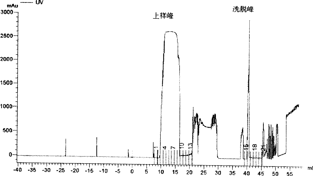
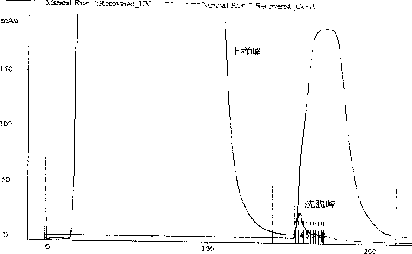
[0129] 3) Load the extractable nuclear antigen solution onto the prepared Jo-1 immunoaffinity chromatographic column, and collect the loading peaks in separate tubes;

[0130] 4) Equilibrate the affinity column with binding buffer for 5-10 column volumes;

[0131] 5) Use elution buffer (3-5M MgCl 2 ) 5-10 column volume elution, separate tubes to collect the elution peak;

[0132] 6) Equilibrate the affinity chromatography column with binding buffer for 5-10 column volume...


PUM


Abstract

The present invention relates to technological process of purifying Jo-1 antigen for diagnosis of multiple myitis/dermatomyositis. The technological process is specific combination of immunoaffinity chromatography and other protein purifying technology. The present invention also relates to the preparation process of Jo-1 immunoaffinity chromatography column, and Jo-1 antigen prepared through the said technological process and with proper biological tissue as material. The present invention also relates to the establishment of high efficiency immunoaffinity chromatography separation condition. The present invention also includes immunoassay kit prepared with purified Jo-1 antigen for clinical application and its application. In addition, the present invention provides one kind of technological way for preparing specific antigen used in detecting other autoimmune diseases.

Description

technical field [0001] The present invention relates to a Jo that can be used for the specific diagnosis of autoimmune diseases polymyositis (PM) / dermatomyositis (DM) and anti-Jo-1 antibody syndrome (Anti-Jo-1 Antibody Syndrome) -A convenient and efficient purification process for the 1 antigen, the Jo-1 antigen prepared by the process method is used to manufacture a clinically applicable polymyositis / dermatomyositis and anti-Jo-1 antibody syndrome specific diagnosis Immunoassay methods, such as the development of dot blot (DB) or enzyme-linked immunosorbent assay (ELISA) diagnostic kits, etc. Background technique [0002] Polymyositis is clinically characterized by symmetrical muscle strength decline in varying degrees at the proximal extremities, and can also involve neck muscles and other skeletal muscles other than facial muscles, and most of them are accompanied by myogenic skeletal muscle enzymes in the blood. Damaged EMG changes, such as creatine kinase (CK), aldolas...

Claims


Application Information

Patent Timeline
Patent Type & Authority Applications(China)
IPC IPC(8): C07K14/47C07K1/22G01N33/557G01N33/535
CPCY02P20/582
Inventor 姚志建唐福林朱材忠陈华彭鲲鹏
Owner SINOGENOMAX
Why Eureka
Try Eureka