Biodegradable dual porous scaffold wrapped with semi-permeable membrane and tissue cell culture using thereof

A technology for organizing cells and biological tissues, applied in the field of scaffolds, can solve the problems of slats, insufficient cell nutrition, and difficulty for cells to grow into the scaffold, etc.

Inactive Publication Date: 2006-03-01
YONSEI UNIVERSITY
View PDF0 Cites 2 Cited by
  • Summary
  • Abstract
  • Description
  • Claims
  • Application Information

AI Technical Summary

Problems solved by technology

[0011] However, traditional fabrication techniques often result in scaffolds with low porosity, uncontrollable pore sizes, and open-pore networks with poor interconnections.
In addition, when tissue cells are implanted and proliferated on the scaffold, the pores on the surface of the scaffold are often blocked, causing difficulties in the preparation of the graft
Therefore, the conventional technology also has the following problems: toxic gas may be generated during the scaffold manufacturing process; slats continue to exist on the scaffold; cells are difficult to grow into the scaffold; and nutrients supplied to the cells are insufficient
Additionally, the growth of histiocytes is not uniform across the scaffold

Method used


Image

  • Biodegradable dual porous scaffold wrapped with semi-permeable membrane and tissue cell culture using thereof
  • Biodegradable dual porous scaffold wrapped with semi-permeable membrane and tissue cell culture using thereof
  • Biodegradable dual porous scaffold wrapped with semi-permeable membrane and tissue cell culture using thereof

Examples


Embodiment 1

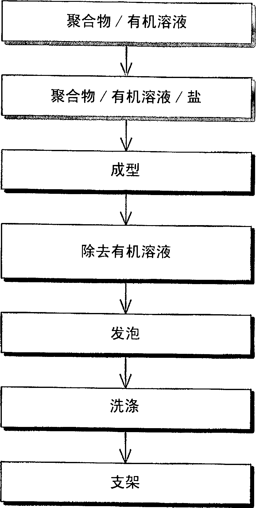
[0084] Embodiment 1: manufacture support

[0085] A mixed solvent was prepared by mixing 90% methylene chloride (Ducksan Chemical Co. Ltd., Korea) and 10% dimethyl sulfoxide (Sigma, USA). PLGA accounting for 10 wt% of the mixed solvent was dissolved in the mixed solvent, wherein PLGA was composed of lactic acid (Sigma, USA) and glycolic acid (Sigma, USA) in a ratio of 75:25, and the molecular weight was 90,000-126,000Da.

[0086] The resulting solution was mixed with ammonium bicarbonate (Junsei Chemical Co. Ltd., Japan) having a particle size of 150-250 μm in a weight ratio of 9:1. The resulting mixture was poured into a cylindrical basin (Dongsung science Co. Ltd, Korea) with a diameter of 100 mm.

[0087] Chloromethane (Ducksan Chemical Co. Ltd., Korea) contained in the mixture charged into the basin (Dongsung Science Co. Ltd., Korea) was partially evaporated to obtain a semi-solidified sample.

[0088] Then, the semi-coagulated sample was immersed in a 30% acetic acid so...

Embodiment 2

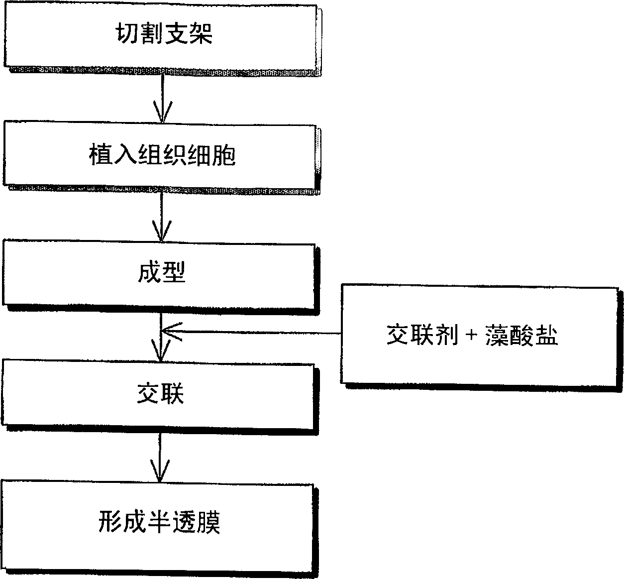
[0093] Embodiment 2: regeneration of biological tissue

[0094] (i) implanting tissue cells on the scaffold

[0095] The stent manufactured in Example 1 was cut into small pieces with a diameter of 2 mm and a thickness of 1 mm. The chondrocytes isolated from rabbits were implanted on the scaffold at a density of 1.0×l05 cells / ml.

[0096] Scaffold pieces were stored at 37°C with 5% CO 2 Incubate in a high humidity environment for about 3 hours to cross-link the scaffold pieces. Then, the scaffold pieces were cultured for 3 days in DMEM (JBI, Korea) containing 4 ml of antibacterial / antifungal solution and FBS (Sigma, USA).

[0097] (ii) Form a semi-permeable membrane on the outer surface of the scaffold and carry out tissue cell proliferation

[0098] 3% sodium alginate solution (Sigma, USA) was mixed with an equal volume of DMEM (JBI, Korea) containing 4 ml of antibacterial / antifungal solution and FBS (Sigma, USA).

[0099] The resulting solution was poured into a 50 ml t...

Embodiment 3

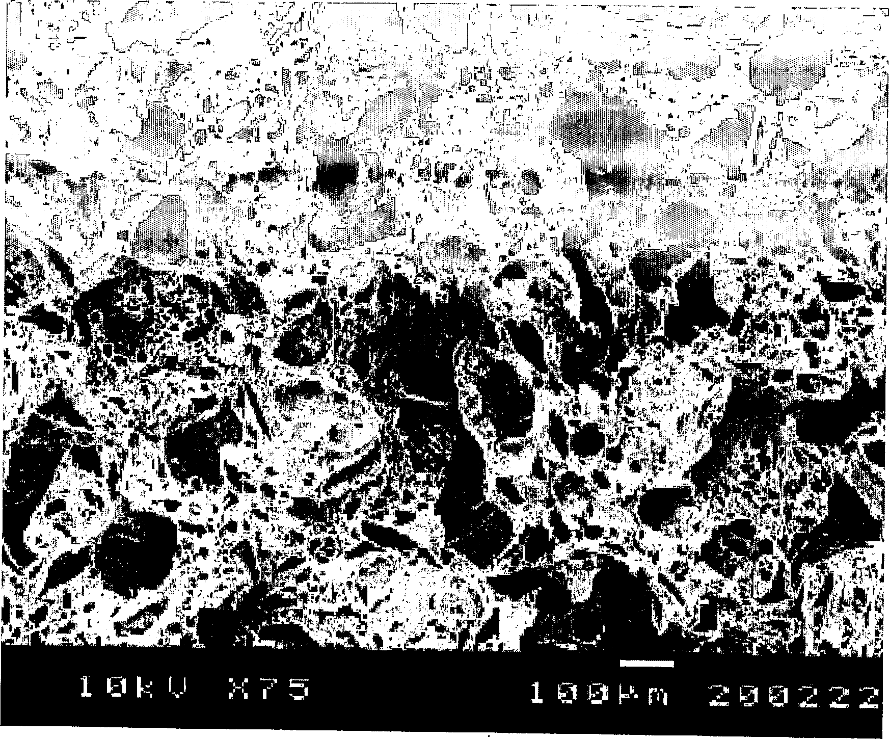
[0104] The gel particle that embodiment 3 makes is at 37 ℃, contains 5% concentration CO 2 In a high-humidity environment, incubate under 90rpm shaker agitation. Change the culture medium every three to four days. On days 7, 14, 21 and 31, the proliferation of chondrocytes was evaluated, and the gel particles were observed with a scanning electron microscope (S-800, Hitachi, Japan), while a flash spectrometer (Luminescence Spectrometer LS50B, PERKIN ELMER, Great British ) fluorescence for quantitative DNA analysis.

[0105] The result is as Figure 6 , 7 and 8 are shown.

[0106] Figure 6 The SEM image of Fig. 1 shows chondrocytes proliferating on the scaffold of the present invention whose outer surface is covered with a semipermeable membrane. Figure 7 The graph of is indicative of the DNA synthesis of chondrocytes grown on the scaffolds of the present invention. Figure 8 The graph of shows the mucopolysaccharide synthesis of chondrocytes grown on the scaffold of t...


PUM

PropertyMeasurementUnit
sizeaaaaaaaaaa
particle diameteraaaaaaaaaa
diameteraaaaaaaaaa

Abstract

Disclosed is a scaffold including a semi-permeable membrane on an outer surface thereof. The present invention also discloses a method of preparing a scaffold covered with a semi- permeable membrane, including loading one or more scaffolds into a mold with a predetermined form and size; and adding a semi-permeable agent and a cross-linking agent to the mold and cross-linking the semi-permeable agent to form the semi-permeable membrane on the outer surface of each of the scaffolds. The scaffold covered with the semi-permeable membrane selectively introduces nutrients into the scaffold by allowing penetration of only external nutrients into the scaffold and excreting metabolic wastes generated by tissue cells to the outside of the scaffold. In addition, the scaffold has the morphology of a biological tissue of interest by cross-linking the small-sized scaffolds, thereby allowing uniform proliferation of tissue cells throughout the whole scaffold.

Description

technical field [0001] The present invention generally relates to a scaffold and a method for preparing biological tissue using the scaffold. More specifically, the present invention relates to a regeneration method of biological tissue. The regeneration method is to use a biodegradable polymer to foam the gas of an effervescent salt to prepare a porous scaffold; divide the scaffold into small pieces; implant tissue cells into onto the stent piece; forming a semipermeable membrane on the outer surface of each stent piece; and cross-linking the stent piece covered by the semipermeable membrane into a predetermined form. [0002] As used herein, the term "scaffold" refers to a porous biodegradable polymeric structure that supports cell growth and migration. Background technique [0003] Normally, cartilage is a tissue that does not naturally regenerate once damaged. To repair damaged cartilage tissue, cartilage substitutes, such as nonabsorbable biomaterials, have been used....

Claims


Application Information

Patent Timeline
Patent Type & Authority Applications(China)
IPC IPC(8): C12N5/00C12N5/06A61K9/00A61L27/20A61L27/56
CPCA61F2002/30766C12N5/0068A61F2002/30762A61L27/20C12N2533/74C12N2533/40A61L27/56C08L5/04C12N11/02C12N5/0602
Inventor 金重贤李惠苑崔星旭
Owner YONSEI UNIVERSITY
Why Eureka
Try Eureka