Particle agglutination detection method and device

A particle and particle suspension technology, applied in the direction of measuring devices, biochemical equipment and methods, microbial measurement/testing, etc., can solve the problems of bulky test tubes, appearance misunderstanding, complexity, etc., to avoid false positive results, easy to use, Effects of Low Margin of Error

Inactive Publication Date: 2006-10-18
INVERNESS SWITZERLAND GMBH
View PDF5 Cites 14 Cited by
  • Summary
  • Abstract
  • Description
  • Claims
  • Application Information

AI Technical Summary

Problems solved by technology

Some drawbacks of this method include: (a) the need to mix for several minutes; (b) subjective visual results; (c) false positive reactions due to drying; and (d) inability to store slides
Some of the pitfalls of tube agglutination include: (a) the need for laboratory equipment, personnel, and environment; (b) subjective visual inspection of the results; (c) incorrect centrifugation speed or time may lead to false-positive or false-negative results, as possible Cell clumps considered immunoagglutinated; (d) no direct recording of results; (e) bulky tubes; and (f) risk of breakage and spillage
Drawbacks are: (a) require laboratory instruments, personnel and environment; b) do not record results directly; (c) bulky test hardware (even though the manufacturer calls the product a "card" it is actually in the holder series of test tubes, and not as flat as a "card"); and (d) complex preparation (inserting gel into narrow test tubes) resulting in high cost
Although the Akers device simplifies blood typing to such an extent that it can be used outside the laboratory (e.g., at the bedside) by non-blood bank professionals, it still has some disadvantages: (a) it requires a relatively large volume of blood; ( b) it requires the operator to record the timing of the steps; (c) it only allows for positive blood typing and not for crossmatch (observing the activity of the recipient's antibodies against the donor's red blood cells); (d) the appearance of the results may Can be misleading: the absence of red in the result window indicates a positive result, while red appears in a negative reaction result; and (e) the result must be observed exactly 1 minute after adding the saline wash
Although these products actually simplify the blood testing procedure to some extent, they are not completely error free and can be complicated for some personnel (Rachel and Plapp, 1990; Migeot et al., 2002)

Method used


Image

  • Particle agglutination detection method and device
  • Particle agglutination detection method and device

Examples


Embodiment approach

[0137] In another embodiment of the invention, a device is provided having multiple test areas (eg, for blood typing of a single sample). The device can be constructed so that the test sample, and optionally the wash, is dispersed throughout all of the test areas in one step. Such an embodiment can be achieved through various design points, such as: (a) covering all test areas with a single piece, preferably hydrophilic mesh; (b) by placing non-absorbent A permanent covering creates a capillary space on the test area.

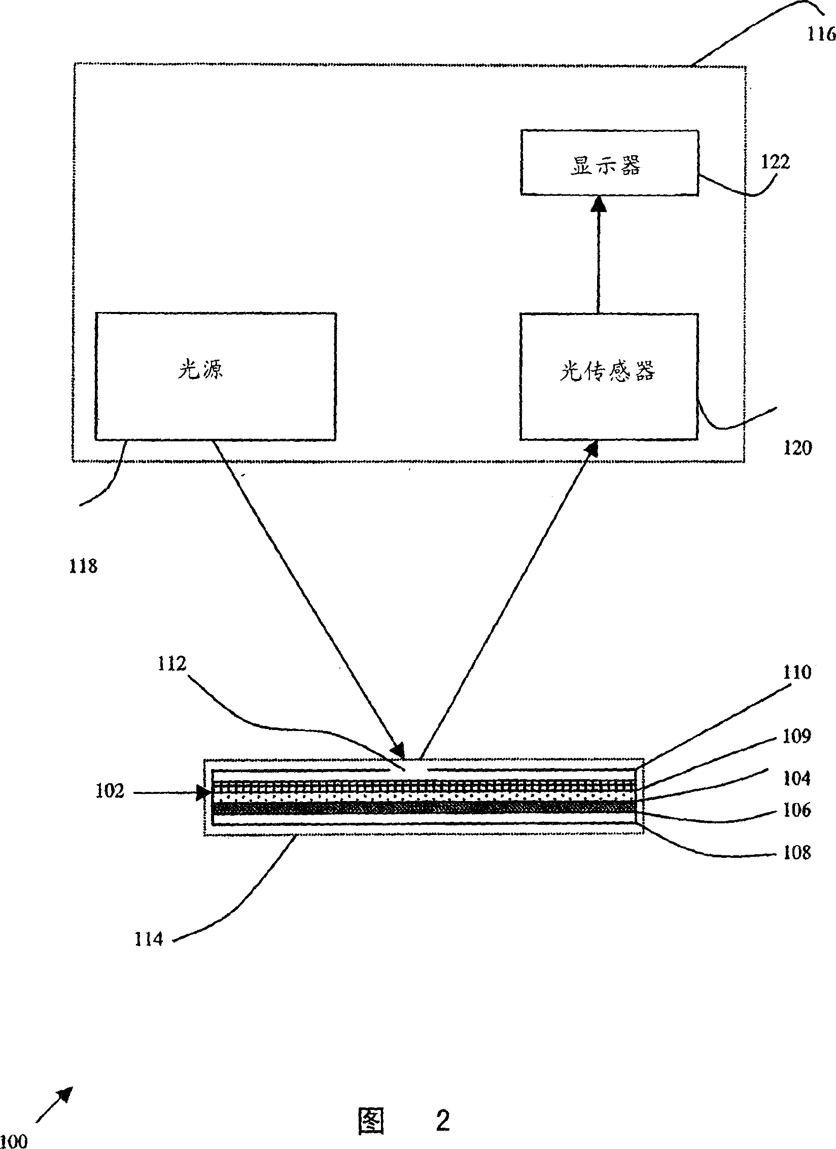
[0138] The device may further include an optional meter 116 that can interpret test results without human visual identification. Meter 116 preferably receives card 102 to enable meter 116 to measure a marker or reporter for detecting agglutination. Optionally, meter 116 is an optical meter and measures the amount of light reflected or scattered from or transmitted through the location where the sample is placed and determines the result accordingly. In its m...

Embodiment

[0142] The following examples are illustrative implementations of the methods and systems described herein. These examples are not intended to be limiting in any way.

[0143] Example 1 - Lotion

[0144] The washes used to form this example were:

[0145] Dulbecco's phosphate buffered saline (PBS) was obtained from Biological Industries, Beit Ha'emek, Israel.

[0146] Solutions were made of PBS diluted 1:1 in water with 4% w / v polyethylene glycol (PEG) 15000-20000 MW (Fluka) and 0.3% w / v dextran sulfate sodium salt (Amersham Biosciences).

[0147] A. Dulbecco's phosphate buffered saline (PBS) with 0.001-0.01% w / v polyoxyethylene-10-tridecyl ether (Sigma).

Embodiment 2

[0148] Embodiment 2-blood type identification

[0149] A 0.5 x 0.5 cm piece of Ahlstrom #142 filter paper was placed on an absorbent pad. 2 μL of whole blood was pipetted into the center of the filter, followed by 2 μL of anti-blood typing reagent (GammaBiologicals Inc., Hosuton, TX, USA). The filter was then washed with a few drops (from a dropper bottle) of washing solution and dried. Alternatively, use 4 mm diameter round Ahlstrom #142 filter paper, 10 μL blood and 10 μL antiblood group reagent, followed by a few drops of Wash Solution C from Example 1.

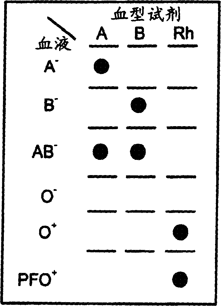
[0150] This test was repeated with multiple blood samples of various blood types (sourced and pre-tested by Central Blood Services, Israel Red Magen David, Tel Hashomer, Israel), each of which was tested with anti-A, anti-B, anti- AB and anti-D (=Rh) blood group reagents were tested. A-blood produces red dots only with anti-A and anti-AB reagents. B-blood was spotted with anti-B and anti-AB reagents. AB-blood reacts w...


PUM


Abstract

A method for detecting and / or observing particle agglutination comprising: placing a volume of particle suspension on a filter constructed to allow passage of individual particles; placing a volume of solution or suspension containing an agglutination reagent on the particle The location of the suspension; optionally placing the washing solution at the location of the agglutinating reagent; and observing the surface of the location for the presence of particles, which presence indicates that particle agglutination has occurred. Also provided are methods for detecting agglutination in general and hemagglutination, eg, for blood typing and cross-matching in particular. The method consists of continuous vertical addition of whole blood, blood typing reagents, and detergent to the filter. In the case of hemagglutination, colored, preferably red or reddish spots appear after washing. Devices and kits based on the present invention are also claimed which facilitate blood typing and cross matching in a non-laboratory environment without the need for laboratory instruments.

Description

field of invention [0001] The present invention relates generally to the detection of receptor-ligand interactions, and more particularly to the detection of blood group antigens and antibodies thereto for blood typing and matching purposes in transfusion medicine. Background of the invention [0002] Particle agglutination is a widely adopted immunological method for detecting and visualizing antigen-antibody interactions due to its simplicity, rapidity and relative sensitivity (Riochet 1993). Cells in general, and erythrocytes in particular, are particles that can be subjected to various methods of agglutination. Thus, erythrocyte agglutination (hemagglutination) is used in blood typing or matching as an important component of the pre-transfusion testing process to detect antigens on their surface and antibodies against such antigens (Rouger 1993; Brecher 2002). The purpose of pre-transfusion testing of blood is to prevent adverse reactions caused by hemagglutination or h...

Claims


Application Information

Patent Timeline
Patent Type & Authority Applications(China)
IPC IPC(8): G01N33/80C12Q1/68G01N33/543
CPCG01N33/80
Inventor G·罗特F·塞缪尔斯F·菲什
Owner INVERNESS SWITZERLAND GMBH
Why Eureka
Try Eureka