

Paper strip for quantitative electrochemical biological test
A quantitative detection and test strip technology, which is applied in biological testing, material electrochemical variables, microbial measurement/inspection, etc., can solve the problems of limited immobilization of biomolecules and affecting detection sensitivity, etc.
- Summary
- Abstract
- Description
- Claims
- Application Information
AI Technical Summary
Problems solved by technology
Method used
Image

Examples
Embodiment 1
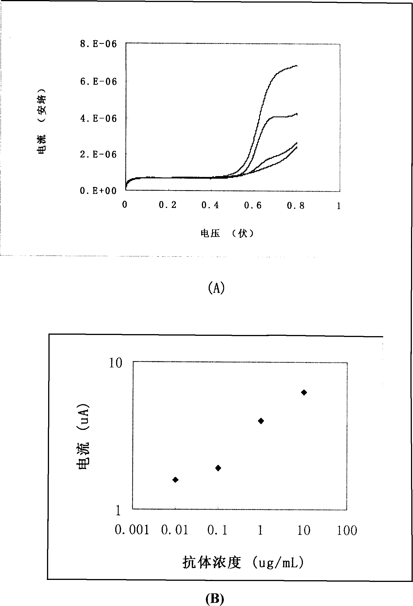
[0016] Example 1: Electrochemical method to detect the immune reaction between mouse IgG / goat anti-mouse IgG on nitrocellulose membrane.
[0017] Take a square nitrocellulose membrane with a side length of 1 cm and soak it overnight in 10 μg / mL mouse IgG (dissolved in 20 mM sodium carbonate / sodium bicarbonate buffer, pH 9.5). After washing, block with 1% BSA for 2 hours. Add different concentrations of alkaline phosphatase-labeled goat anti-mouse antibody, and shake at room temperature for 1 hour. After cleaning, the membrane was placed on the screen-printed electrode sheet and phosphate phenol was added. After waiting for 5 minutes, start the cyclic voltammetry detection program of the electrochemical workstation, and record the current signal. get as figure 2 The results shown show that the detected current signal has a linear relationship with the concentration of the enzyme-labeled antibody within a certain range, and the lowest level of 0.1 μg / mL (0.7nM) antibody can ...
Embodiment 2
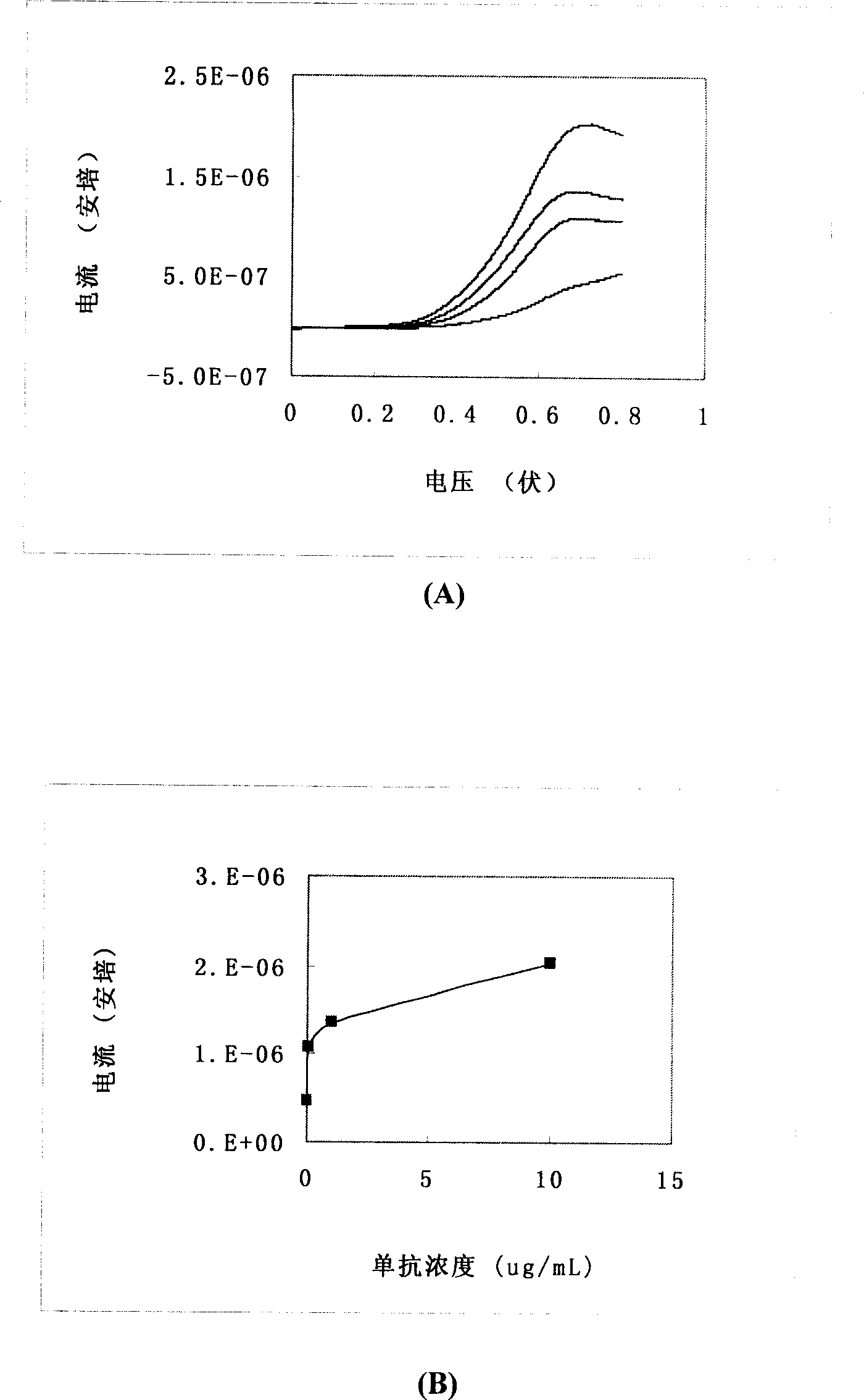
[0018] Example 2: Electrochemical method for detection of immune reaction between estradiol antigen / antibody on nitrocellulose membrane.
[0019] Take a square nitrocellulose membrane with a side length of 1 cm and soak it overnight in 10 μg / mL estradiol antigen (dissolved in 20 mM sodium carbonate / sodium bicarbonate buffer, pH 9.5). After washing, block with 1% BSA for 1 hour. Different concentrations of estradiol mouse monoclonal antibody were added, and the reaction was shaken at room temperature for 1 hour. After washing, add alkaline phosphatase-labeled goat anti-mouse secondary antibody diluted 1000 times and react with shaking at room temperature for 1 hour. After cleaning, the membrane was placed on the screen-printed electrode sheet and phosphate phenol was added. After waiting for 5 minutes, start the cyclic voltammetry detection program of the electrochemical workstation, and record the current signal. get as image 3 The results shown indicate that the detected...
Embodiment 3
[0020] Example 3: Electrochemical method to detect the competitive immune response of estradiol on nitrocellulose membrane.
[0021] Take a square nitrocellulose membrane with a side length of 1 cm and soak it overnight in 10 μg / mL estradiol antigen (dissolved in 20 mM sodium carbonate / sodium bicarbonate buffer, pH 9.5). After washing, block with 1% BSA for 1 hour. Add different concentrations of estradiol and 0.1 μg / mL estradiol mouse monoclonal antibody, and shake at room temperature for 1 hour. After washing, add alkaline phosphatase-labeled goat anti-mouse secondary antibody diluted 1000 times and react with shaking at room temperature for 1 hour. After cleaning, the membrane was placed on the screen-printed electrode sheet and phosphate phenol was added. After waiting for 5 minutes, start the cyclic voltammetry detection program of the electrochemical workstation, and record the current signal. get as Figure 4 The results shown indicate that the detected current sign...
PUM
 Abstract
Description
Claims
Application Information