
Magnetic resonance maps for analyzing tissue

a magnetic resonance map and tissue technology, applied in the field of magnetic resonance map, can solve the problems of limiting the validity of a progression free survival end point, premature discontinuation of effective adjuvant therapy, and inability to reliably distinguish between tumor progression and treatment effects of conventional magnetic resonance map, and achieve high resolution and depiction. high

Active Publication Date: 2019-04-30
RAMOT AT TEL AVIV UNIV LTD +1
View PDF51 Cites 3 Cited by
  • Summary
  • Abstract
  • Description
  • Claims
  • Application Information

AI Technical Summary

Benefits of technology

[0048]In the maps of some embodiments of the present invention, progression is reflected by a significant increase in the fast component of the enhancing lesion while pseudoprogression is reflected by an increase in the slow component with no significant increase in the fast component. Thus, using the maps of the present embodiments in routine MRI follow-up may aid the physician in determining progression versus pseudoprogression in patients presenting an increase in the enhancing lesion on T1-MRI. An increase in the fast component volume, suggesting progression, implies that a change in the current therapy should be employed. No significant increase in the fast volume component, suggesting pseudoprogression, implies that the patient is responding to the current therapy and thus continuation is preferred, if possible.
[0050]The ability of the maps of the present embodiments to depict morphologically active tumor regions with high resolution can be applied for optimizing radiation treatment planning by localizing the treatment to the fast / blue regions in the maps. This methodology can optionally and preferably be applied in post surgery scenario to allow differentiation between post surgical changes and tumor remnants thus allowing depicting and treating residual tumor post surgery.
[0052]The maps of the present embodiments may also be applied for guiding stereotactic biopsies for molecular classification of the tumor. In this case the maps may aid in preventing the acquisition of biopsies with a high amount of necrosis or out of the infiltration zone with ‘contamination’ of the specimen by normal brain tissue, both potentially leading to false-negative results.
[0055]The high sensitivity of the maps of the present embodiments can provide additional information for delineation of the tumor borders as well as for targeting stereotactic biopsies. The results presented herein demonstrate the applicability of the technique of the present embodiments in the daily clinical scenario. The ability to clearly differentiate tumor from non-tumoral tissues provides the physician with a clear understanding of the patient current situation thus enabling improved patient management.

Problems solved by technology

This treatment related effect has implications for patient management and may result in premature discontinuation of effective adjuvant therapy.
This limits the validity of a progression free survival end point unless tissue-based confirmation of tumor progression is obtained.
Treatment decision, as whether to operate on a patient with radiographic deterioration, continue chemoradiation or change to another non-surgical treatment is a day to day struggle involving interdisciplinary teams of neurosurgeons, neuro-oncologists and neuro-radiologists who are often unable to reach a unanimous interpretation of the patient status.
For glioblastoma patients treated by radiation and TMZ, conventional MRI is unable to provide a reliable distinction between tumor progression and treatment effects (also referred to as pseudoprogression).
For glioblastoma patients conventional MRI is unable to reliable depict the tumor or pseudoprogression after treatment with Avastin.
There is limited, but increasing evidence that PET with amino acid tracers can discriminate treatment-related necrosis from tumor recurrence.
Unfortunately, there was significant overlap between the two groups.
The prognosis of patients diagnosed with brain metastases is generally poor.
Among the reported complications of SRS is radiation-induced necrosis which, similarly to pseudoprogression, can be difficult to differentiate both clinically and radiologically from recurrent tumor at the treatment site.

Method used


Image

  • Magnetic resonance maps for analyzing tissue
  • Magnetic resonance maps for analyzing tissue
  • Magnetic resonance maps for analyzing tissue

Examples


example 1

[0142]Patients and Treatment:

[0143]Twelve patients with histologically confirmed glioblastoma (World Health Organization [WHO] grade IV astrocytoma) were recruited. Eleven with newly diagnosed GBM and one patient with secondary GBM, two years after resection of an Anaplastic Astrocytoma (WHO grade III tumor). All patients received the standard radiotherapy of 60 Gy in thirty daily fractions, five days a week, with concomitant daily temozolomide of 75 mg / m2 for forty two days. Chemoradiation was followed by six cycles of adjuvant temozolomide of 150-200 mg / m2 daily for five days every twenty eight days. Three patients were recruited prior to surgery and nine after completion of the chemoradiation treatment. The mean age was 50.7±3.7 with a range of 27-71. Nine of the twelve patients were men.

[0144]Inclusion criteria included patients with WHO performance status of 2 or less and adequate hematologic, renal, and hepatic function. Exclusion criteria included contraindications to MRI and...

example 2

[0189]An application of the present embodiments comprises high resolution depiction of brain tumors after treatment with anti-angiogenic treatments and studying the mechanism of action and response patterns of such drugs.

[0190]Standard Treatment:

[0191]Glioblastoma multiforme (GBM) is the most common and most aggressive type of high grade glioma and is associated with a median survival of less than 15 months. In 2005, a randomized phase 3 trial demonstrated that the addition of temozolomide (TMZ) to adjuvant radiation therapy was associated with an improvement in the median survival of patients with newly diagnosed GBM from 12.1 months to 14.6 months [1]. This treatment regimen is currently the standard of therapy for newly diagnosed GBM, and is commonly referred to as the “Stupp protocol”.

[0192]Anti-Angiogenic Treatments:

[0193]Nearly all GBM recur after initial therapy, and most patients do not survive beyond 1 year after the diagnosis of recurrent disease. Because re-operation and ...

example 3

[0271]The present embodiments were used in high resolution depiction of brain metastases and other brain disorders after treatment with radiosurgery or other radiation-based treatments for differentiation between tumor progression and radiation necrosis, and in application of high resolution delayed extravasation MRI for differentiation between progression of brain Space occupying lesion (SOL) and treatment effects following radio-surgical treatment.

[0272]Patients and Treatment:

[0273]32 patients are used in the study, 15-75 years of age, post radio surgical treatment of 18-20 Gy targeting a brain SOL with follow up MRIs that may be related to tumor progress or treatment effects. The patients are divided into the following 4 groups:

[0274](1) 6-8 patients with suspected SOLs (brain metastases, AVMs, primary brain tumors or meningiomas) progression,

[0275](2) a control group of the same number (6-8) of patients with similar pathologies who underwent a radio surgical treatment within a s...


PUM


Abstract

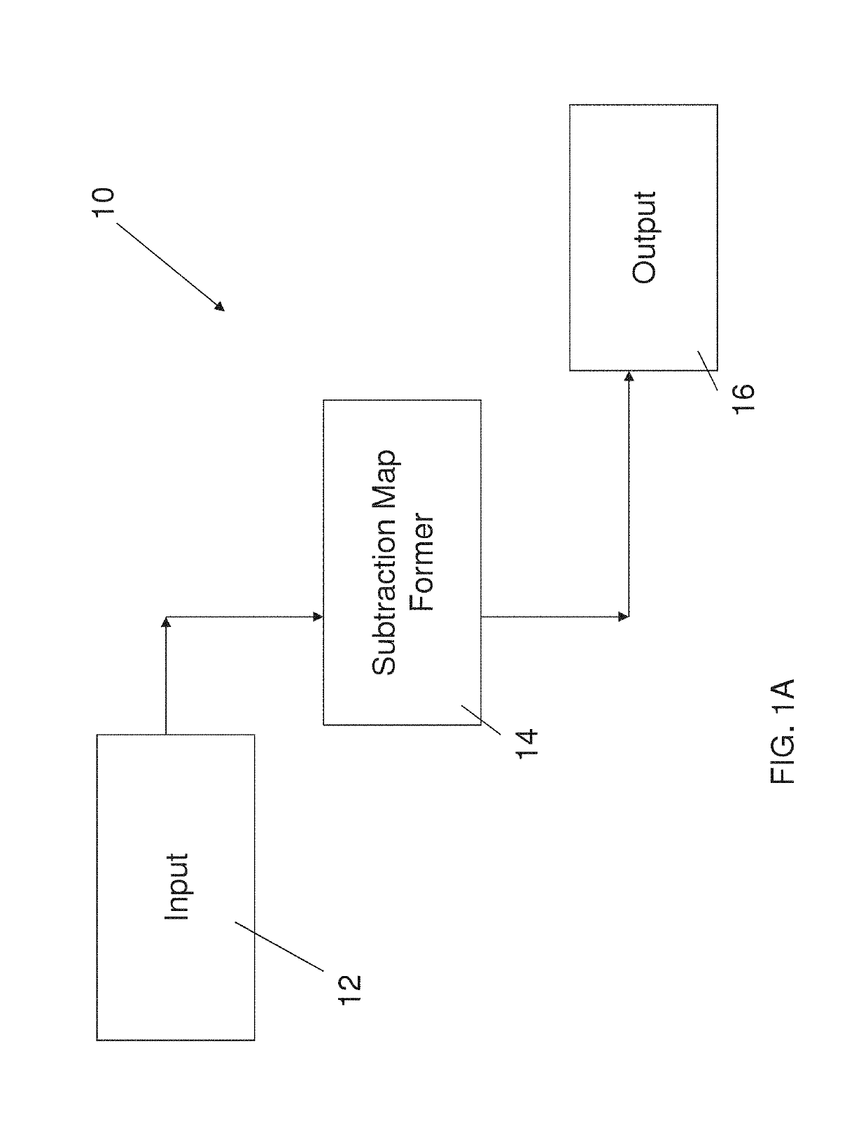
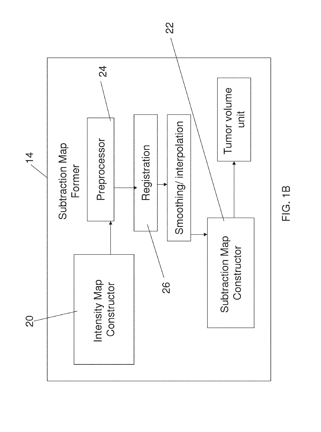
Apparatus for analyzing brain MRI, is disclosed. The apparatus comprises an input for receiving a first and a second MRI scans at the beginning and end of a predetermined time interval post contrast administration; a subtraction map former for forming a subtraction map from said first and said second MRI scans by analyzing said scans to distinguish between two primary populations, a slow population, in which contrast clearance from the tissue is slower than contrast accumulation, and a fast population in which clearance is faster than accumulation; and an output to provide an indication of distribution of said two primary populations, wherein said predetermined time period is at least twenty minutes.

Description

RELATED APPLICATIONS[0001]This application is a National Phase of PCT Patent Application No. PCT / IB2012 / 055703 having International filing date of Oct. 18, 2012, which claims the benefit of priority under 35 USC § 119(e) of U.S. Provisional Patent Application No. 61 / 548,737 filed on Oct. 19, 2011. The contents of the above applications are all incorporated by reference as if fully set forth herein in their entirety.FIELD AND BACKGROUND OF THE INVENTION[0002]The present invention relates to method and apparatus for producing magnetic resonance (MR) maps and, more particularly, but not exclusively to using such maps to identify and study tumors, particularly but not necessarily brain tumors.[0003]Gliomas are the most common malignant primary brain tumors in adults, with an annual incidence of 4-5 per 100,000 people (Central Brain Tumor Registry; Wen and Kesari, 2008).[0004]The standard of care for glioblastoma includes chemotherapy during and after radiotherapy. The use of temozolomid...

Claims


Application Information

Patent Timeline
Patent Type & Authority Patents(United States)
IPC IPC(8): G06K9/62A61B5/00A61B5/055G06T7/00G16H50/50
CPCG06K9/6202A61B5/0042A61B5/055G06T7/0012G16H50/50A61B5/0037A61B2576/026
Inventor ZACH, LEORGUEZ, DAVIDLAST, DAVIDDANIELS, DIANNEMARDOR, YAEL
Owner RAMOT AT TEL AVIV UNIV LTD
Learn More