

Treatment of damaged nerve with PTEN inhibitor
a nerve and pten inhibitor technology, applied in the field of pten inhibitor treatment of damaged nerves, can solve the problems that the exclusion of extracellular inhibitory molecules secreted upon nerve injury only triggers very limited axon regeneration in vivo, and achieves the effect of growing, proliferating or enhancing the activity of nerve cells
- Summary
- Abstract
- Description
- Claims
- Application Information
AI Technical Summary
Benefits of technology
Problems solved by technology
Method used
Image

Examples
example 1
Example 1.1
[0101]Rat adrenal medullary PC12 pheochromocytoma neuronal cell was purchased from ATCC (Manassas, Va.). Cell culture materials including Dulbecco's modified Eagle's medium (DMEM), fetal bovine serum (FBS) and horse serum were purchased from Mediatech Inc. (Manassas, Va.). 2.5 S Nerve growth factor was purchased from BD Biosciences, Inc. (Bedford, Mass. 01730). TUJ-1 monoclonal rabbit antibody against neuronal class III β-tubulin was purchased from Covance Inc. (Gaithersburg, Md.). Monoclonal mouse antibody against acetylated α-Tubulin was purchased from Santa Cruz Biotech Inc. (Santa Cruz, Calif.). Goat serum, Texas Red® Goat Anti-Rabbit IgG antibody, Alexa Fluor® 488 Goat anti-Mouse IgG antibody, 4′,6-Diamidino-2-Phenylindole, Dilactate (DAPI) and AlamarBlue® were purchased from Molecular Probes-Invitrogen (Eugene, Oreg.). Nocodazole was purchased from Sigma-Aldrich (St. Louis, Mo.). Neurite Outgrowth Assay Kit was purchased from Millipore (Bille...
example 1.2
ign
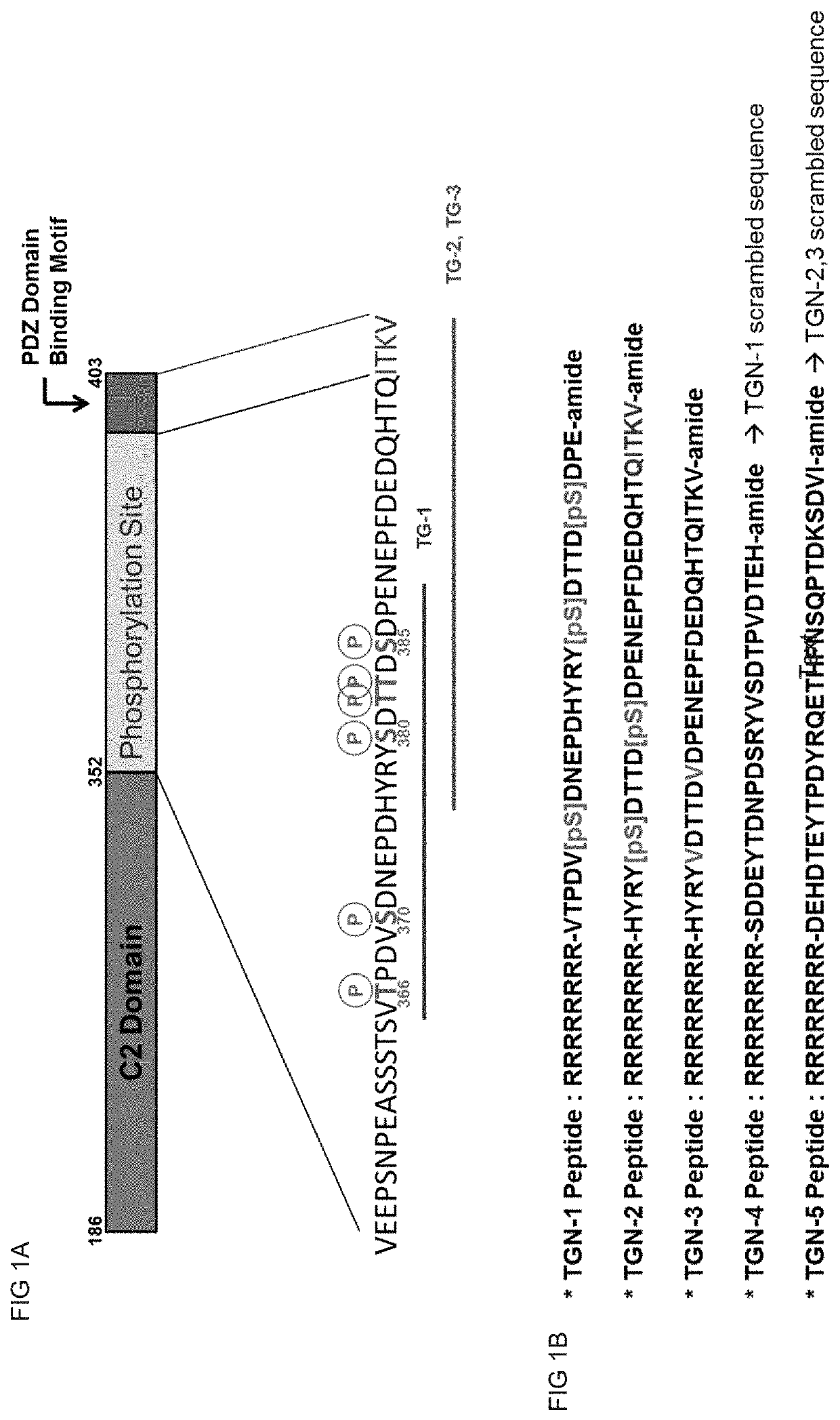
[0102]TGN peptides as potential PTEN inhibitor were designed using PTEN C-terminal region (AA352˜403) as template. All TGN peptides include PTD (peptide transfer domain) sequence (RRRRRRRR) at their N-terminal end to increase membrane permeability. TGN-1 peptide has 32 amino acids with three phosphorylated Serine residues (MW=4244.18 Da, sequence: RRRRRRRR-VTPDVpSDNEPDHYRYpSDTTDpSDPE-amide (SEQ ID NO:4), pS=phosphorylated Serine). TGN-2 peptide has 36 amino acids with two phosphorylated Serine residues (MW=4776.28 Da, sequence: HYRYpSDTTDpSDPENEPFDEDQHTQITKV-amide (SEQ ID NO:6), pS=phosphorylated Serine). TGN-3 peptide has the same amino acid sequence as TGN-2 peptide but no residue is modified and two Serine residues were substituted to Valine (MW=4640.99 Da, sequence: RRRRRRRR-HYRYVDTTDVDPENEPFDEDQHTQITKV-amide (SEQ ID NO:8)). TGN-4 peptide was designed as a scrambled peptide of TGN-1 peptide (MW=4004.19 Da, sequence=RRRRRRRR-SDDEYTDNPDSRYVSDTPVDTEH-amide (SEQ ID NO:10)) and TG...
example 1.3
EN Activity Assay
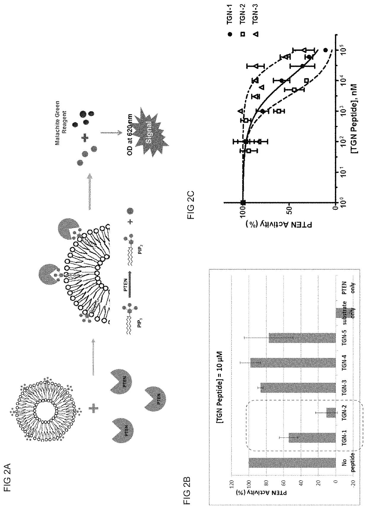
[0103]In vitro PTEN activity assay was designed to check PTEN lipid phosphatase activity to convert phosphatidylinositol triphosphate (PIP3) to phosphatidylinositol diphosphate (PIP2) and produce phosphate ion (Pi). 1,2-dioctanoyl-sn-glycero-3-phospho-(1′-myo-inositol-3,4,5-triphosphate) (C8-PIP3) was used as PTEN substrate and prepared as lipid vesicle (liposome) with other phospholipids because PTEN as lipid phosphatase is an interfacial enzyme. For liposome preparation, C8-PIP3, DOPS (1,2-dioeloyl-sn-glycero-phosphoserine) and DOPC (1,2-dioeloyl-sn-glycero-phosphocholine) were mixed together with 800 μL of liposome buffer (50 mM Tris, 100 mM NaCl, 10 mM MgCl2, 5 mM DTT, pH=8.0) to final concentration of 0.1 mM of C8-PIP3, 0.25 mM DOPS and 0.25 mM DOPC. The lipid mixture was then sonicated at 4° C. for 30 min to produce liposome. After sonication, the liposome solution was briefly centrifuged to remove remaining lipids.
[0104]For PTEN activity assay, 20 ng of recom...
PUM
| Property | Measurement | Unit |
|---|---|---|
| volume | aaaaa | aaaaa |
| concentration | aaaaa | aaaaa |
| temperature | aaaaa | aaaaa |
Abstract
Description
Claims
Application Information