In vivo detection of EGFR mutation in glioblastoma via MRI signature consistent with deep peritumoral infiltration

a technology of glioblastoma and egfr, which is applied in the field of in vivo detection of egfr mutation in glioblastoma via mri signature consistent with deep peritumoral infiltration, can solve the problems of difficult to determine the heterogeneous molecular characteristics of patients, the overall survival rate of gbm patients has not been substantial, and the tumor size is reduced. , the effect of reducing the presence and ,

Active Publication Date: 2021-08-31
THE TRUSTEES OF THE UNIV OF PENNSYLVANIA
View PDF5 Cites 0 Cited by
  • Summary
  • Abstract
  • Description
  • Claims
  • Application Information

AI Technical Summary

Problems solved by technology

Although these treatment options have improved over the years, there has not been any substantial improvement in the overall survival rates of GBM patients.
Determining the highly heterogeneous molecular characteristics in patients diagnosed with GBM is challenging, as conventional options are based on analysis of tissue, which is invasive and, most importantly, fails to capture the spatial heterogeneity of gene expression, as it is based on an analysis of a localized tissue sample.
Neuro-Oncology 17, 907-909 (2015)] of molecular alterations within the GBM tumor that give rise to sampling error (i.e., analysis of single tissue specimen is not sufficient for determining the dominant mutant expression).
Furthermore, the invasive nature of repeated biopsies makes it nearly impossible to evaluate the dynamic equilibrium of mutations and molecular characteristics that occur during the course of treatment, hence adapt the treatment accordingly.
Patient stratification and selection for treatment is limited for the same reason.
In other cases, the biopsy (or resection) of the tumor might not always be possible, such as in cases of deep-seated tumors, in which there is no sufficient sample size for histopathological analysis.
Finally, molecular testing may be unavailable in certain clinical settings due to cost or equipment availability.
Science 344, 1396-1401 (2014)]; and hence, a single tissue might not be sufficient for determining the dominant expression of the mutant.
This, combined with the fact that repeated biopsies are not considered due to their invasive nature, makes it nearly impossible to evaluate the expression dynamics of molecular characteristics during treatment, and hence, to adapt treatment accordingly.
Patient stratification and selection into treatments is limited for the same reason.
Third, the biopsy (or resection) of the tumor might not always be possible, such as in cases of deep-seated tumors, where there is insufficient sample size for histopathological assessment and analysis.
Finally, retrieval of such molecular characteristics may be unavailable in certain clinical settings, as it requires costly and not widely-available equipment for tissue-based genetic testing, especially when such testing must be performed repeatedly for treatment monitoring.
Despite the fact that more than 90% of tumor recurrences occur in edema due to the highly infiltrative nature of GBM, there is limited research focused in the assessment of this region and its microenvironment [J.-M.
Existing healthy / normal blood vessels can provide oxygen and nutrient supplies sufficient for tumors in early stages, but not enough for larger tumors.
The obtained results suggest that the discrimination of the EGFRvIII mutation status, which is critical for personalized treatment decisions and response evaluation, can be achieved based solely on assessing the peritumoral heterogeneity on in vivo perfusion imaging data, whilst obviating costly and not widely-available tissue-based genetic testing.

Method used


Image

  • In vivo detection of EGFR mutation in glioblastoma via MRI signature consistent with deep peritumoral infiltration
  • In vivo detection of EGFR mutation in glioblastoma via MRI signature consistent with deep peritumoral infiltration
  • In vivo detection of EGFR mutation in glioblastoma via MRI signature consistent with deep peritumoral infiltration

Examples


embodiments

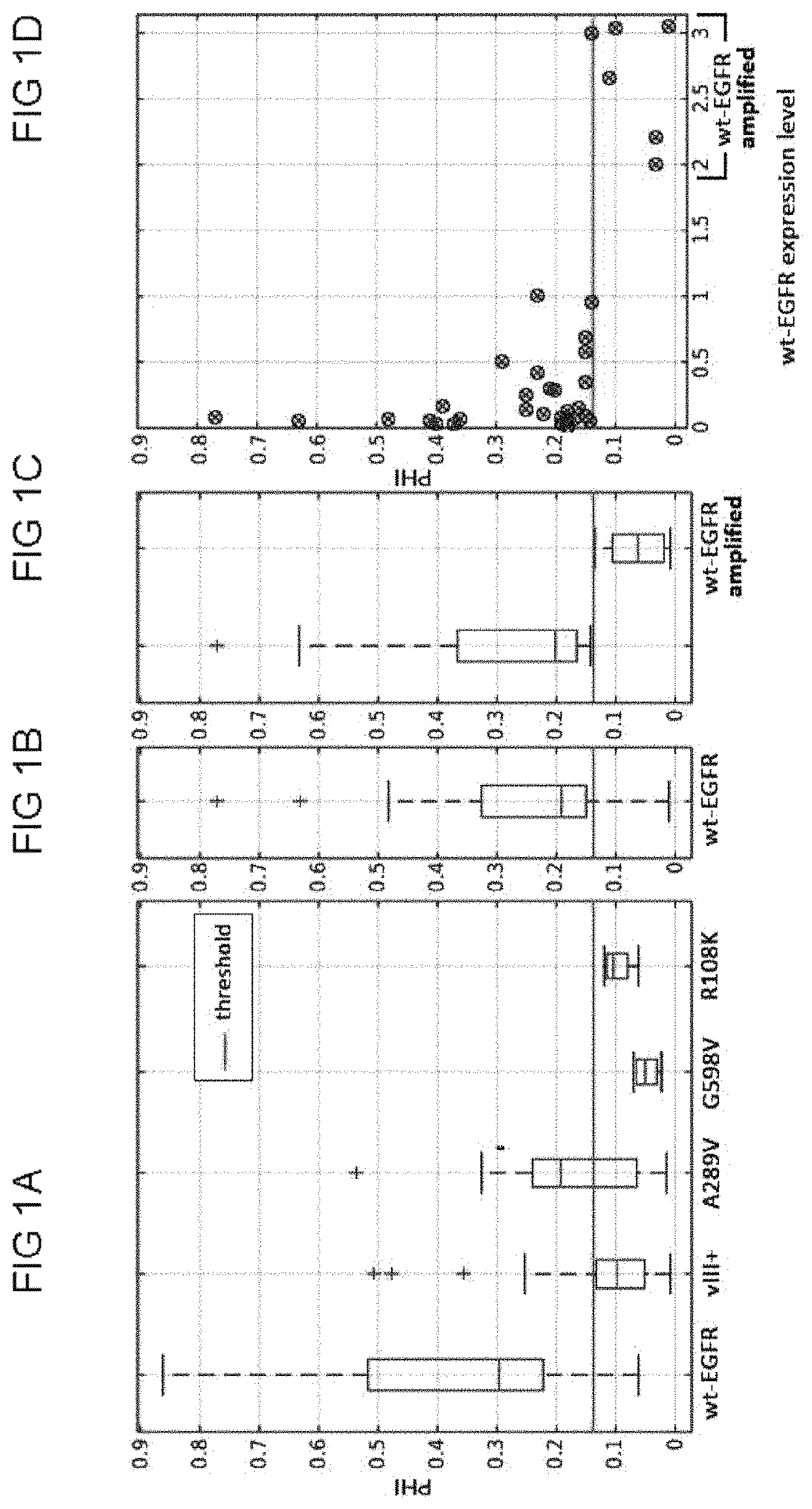
[0053]Embodiments disclosed herein use EGFR mutants as a target for cancer therapy. For instance, EGFR events can be identified to drive one particular molecular subtype of GBM, namely classical GBM, and corresponding “abnormalities” in the EGFR expression may be associated with predicting survival rate and with directing response and treatment toward aggressive therapies.

[0054]Conventionally, EGFR mutant status is determined based on invasive tissue-based approaches, which include immunohistochemistry and next generation sequencing. These processes are hindered by numerous intrinsic and extrinsic factors. First, the expression of molecular characteristics in GBMs is spatially heterogeneous [A. Sottoriva, et al. Intratumor heterogeneity in human glioblastoma reflects cancer evolutionary dynamics. Proc Natl Acad Sci USA 110, 4009-4014 (2013); A. P. Patel, et al. Single-cell RNA-seq highlights intratumoral heterogeneity in primary glioblastoma. Science 344, 1396-1401 (2014)]; and henc...

example

[0092]A specific example of use of the above disclosed method is provided below. This method is not a limitation on the invention.

[0093]Study Design:

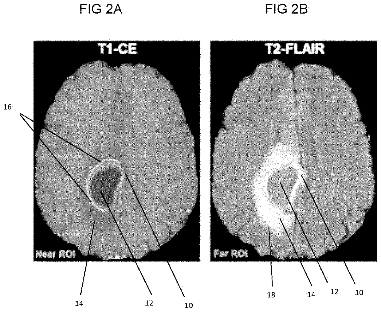
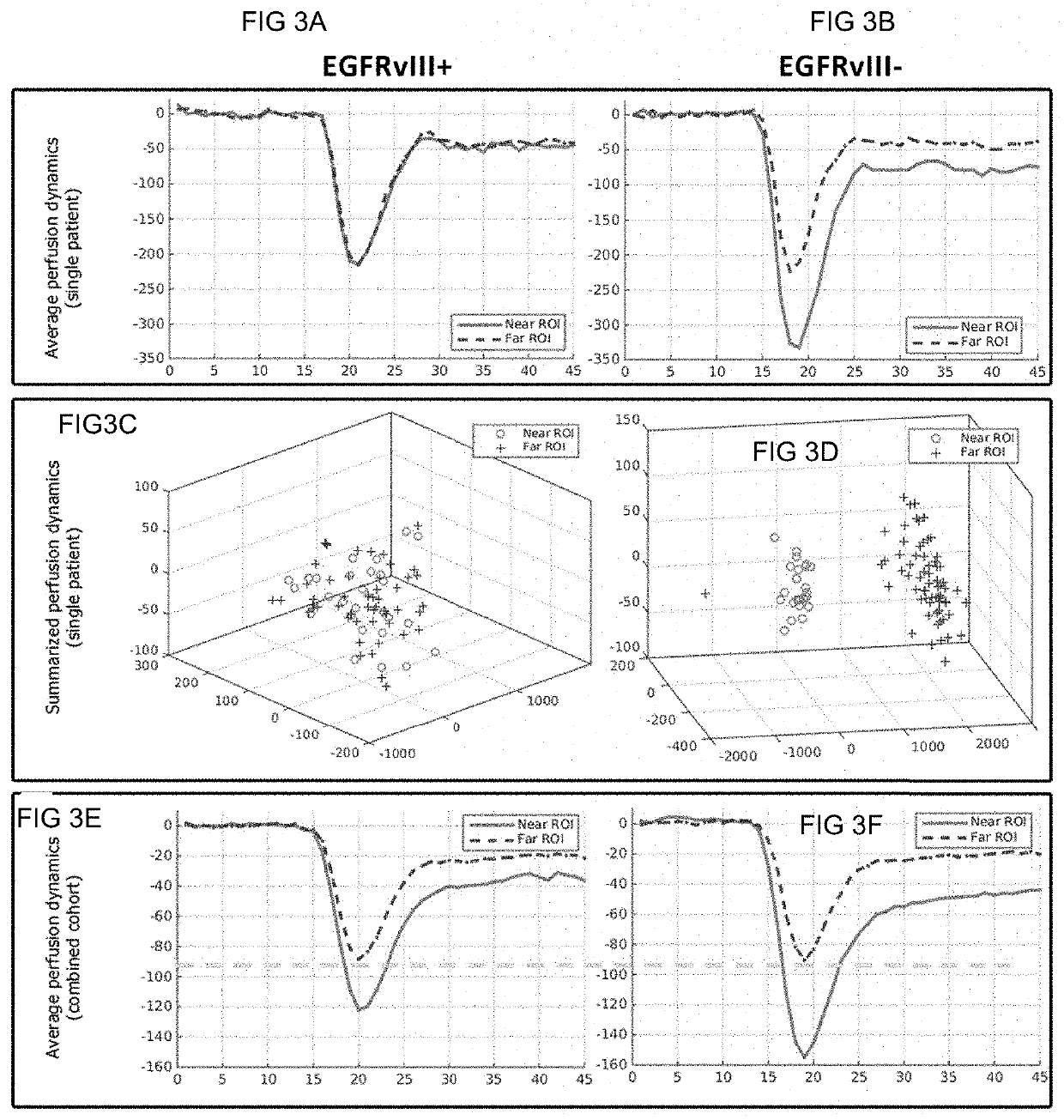
[0094]The data used for this example comprises preoperative multi-parametric MRI data from a retrospective cohort of 142 patients (80 males, 62 females) with de novo GBM. The inclusion criteria for these patients comprised; a) the diagnosis of de novo GBM based on pathology, b) EGFRvIII mutation status based on next generation sequencing, and c) availability of contrast-enhanced T1-weighted, T2-FLAIR, and DSC MRI image volumes. Tissue specimens of these patients were obtained by surgical resection and tested for the status of the EGFRvIII mutation. The patient's mean and median age in years was 59.82 and 60.95, respectively (range: 18.65-86.95). The population of 142 patients was distinguished in 100 (70.42%) with EGFRvIII-negative (57 males, 43 females) and 42 (29.58%) with EGFRvIII-positive (23 males, 19 females) status, which is cons...


PUM


Abstract

A method, including a computer-implemented method, is provided for in vivo detection of epidermal growth factor receptor (EGFR) mutation status within peritumoral edematous tissue of a patient. The method includes performing quantitative pattern analysis of magnetic resonance imaging (MRI) data corresponding to MRI of in vivo peritumoral edematous tissue to determine a level of spatial heterogeneity or similarity within the in vivo peritumoral edematous tissue. EGFR mutation status is assigned as one of negative or positive based on the level of spatial heterogeneity or similarity determined. A non-transitory computer-readable storage medium and a system are also provided.

Description

CROSS-REFERENCE TO RELATED APPLICATIONS[0001]This application claims the benefit of U.S. Provisional Patent Application No. 62 / 484,034, filed Apr. 11, 2017 and U.S. Provisional Patent Application No. 62 / 325,764, filed Apr. 21, 2016, both of which are hereby incorporated in their entireties.STATEMENT OF FEDERALLY SPONSORED RESEARCH[0002]This invention was made with government support from grant R01-NS042645 awarded by the National Institutes of Health (NIH) and grant U24-CA189523 awarded by the NIH. The US government has certain rights in this invention.BACKGROUND[0003]Glioblastoma (GBM) is the most common and aggressive primary malignant adult tumor of the central nervous system. GBM may be located anywhere in the brain or spinal cord, but is typically found in the cerebral hemispheres of the brain. According to the American Brain Tumor Association, GBMs represent about 15.4% of all primary brain tumors and about 60-75% of all astrocytomas [American Brain Tumor Association, Glioblas...

Claims


Application Information

Patent Timeline
Patent Type & Authority Patents(United States)
IPC IPC(8): G06T7/00
CPCG06T7/0014G06T7/0012G06T2207/10088G06T2207/20076G06T2207/20081G06T2207/30016G06T2207/30024G06T2207/30096
Inventor O'ROURKE, DONALDBAKAS, SPYRIDONDAVATZIKOS, CHRISTOS
Owner THE TRUSTEES OF THE UNIV OF PENNSYLVANIA
Why Eureka
Try Eureka