Detection device having capture region and detection region

a detection device and detection region technology, applied in biochemistry apparatus and processes, laboratory glassware, instruments, etc., can solve the problems of cervical cancer development, high labor intensity, and inability to provide established laboratory and sample transportation infrastructure, and achieve simple, rapid molecular diagnostic platform, and immediate visual readout

Active Publication Date: 2021-10-05
TRUSTEES OF BOSTON UNIV
View PDF18 Cites 0 Cited by
  • Summary
  • Abstract
  • Description
  • Claims
  • Application Information

AI Technical Summary

Benefits of technology

[0006]To address this need for easy-to-use, economical diagnostic tools, we have developed a “paperfluidic chip,” i.e., a device made of inexpensive materials (e.g., paper and adhesive film) that serves as a platform combining nucleic acid extraction, amplification (e.g., isothermal loop-mediated amplification), and / or detection (e.g., lateral flow detection) via, e.g., immunochromatographic strips that enable immediate visual readout. This low-cost, portable, and disposable device provides a simple, rapid molecular diagnostic platform for point-of-care detection of nucleic acids, of any source. We developed a fully integrated, on-chip, sample-to-answer assay platform for the detection of various biomarkers directly from patient specimens in under an hour. This novel diagnostic platform could overcome many barriers associated with a variety of settings. For example, the device provides increased access to screening for diseases and early detection for patients in limited-resource settings, improving quality of life and reducing mortality from late detection. The device can be used in low-resource settings to reach patients who may otherwise go undiagnosed or be incorrectly diagnosed due to insufficient resources and personnel. In addition, the device offers an efficient alternative to conventional diagnostic procedures in the context of, e.g., over-the-counter home testing, pharmacy based testing, medical offices, field testing, or emergency rooms.
[0007]In one aspect, the invention features a detection device including a capture region for binding a target in a sample and a waste region that is connected to the capture region and that can absorb a non-target fraction of the sample. The device can further include a detection region separated from the capture region by a removable barrier (e.g., tab) that, when present, prevents transfer of liquid between the capture region and the detection region. The device can also include a non-absorbent housing that prevents moisture transfer (e.g., evaporation, leakage, or transfer between other device components (e.g., between the detection region and the capture region). The capture region typically includes a porous membrane (e.g., polyethersulfone (PES), glass fiber, cellulose paper, polycarbonate membrane, or other polymer or natural material-based porous membrane).
[0008]In some embodiments, the housing of the device can include a film (e.g., an adhesive film). In some embodiments, the film can include the removable tab separating the detection region from the capture region. In some embodiments, the film is perforated for tearing or folding. In some embodiments, the waste region can be disconnected from the capture region by tearing (e.g., along one or more of the perforations). In some embodiments, the removable tab can similarly be disconnected from the device by tearing, e.g., as part of the removal of the waste region or by folding at the perforation to disrupt contact between the capture region and the waste region. In some embodiments, the device also includes a protective flap (e.g., as part of the film) that can be folded onto the capture region to prevent moisture loss (e.g., by evaporation or leakage). In some embodiments, the film is cut from a single planar sheet. In some embodiments, the detection region and capture region are housed in separate plastic casings that can be moved in and out of contact with each other by turning, sliding, or “clicking” the plastic housings into place.

Problems solved by technology

Infectious diseases disproportionately affect the developing world where standard molecular diagnostic approaches requiring expensive equipment, highly skilled technicians, and established laboratory and sample transportation infrastructure are unavailable.
In some women, however, the infection becomes persistent and may lead to the development of cervical cancer.
While these results validate the use of HPV DNA testing, a significant drawback is the high cost and the need for sophisticated laboratory equipment.
Furthermore, current HPV DNA tests still require highly trained laboratory personnel and incur turnaround times of hours to days, depending on how far the sample has to travel to a central laboratory (Villa et al, Int. J. of Gynecology &Obstetrics, 2006, 94, S71-S80).
These requirements result in expensive equipment and costly disposables unsuitable for use in limited resource settings.
Immunoassay-based rapid diagnostic tests (RDTs) offer a faster, lower-cost solution for resource-limited settings, but they suffer from low test sensitivities and specificities, commonly resulting in false negative and / or false positive detection.

Method used


Image

  • Detection device having capture region and detection region
  • Detection device having capture region and detection region
  • Detection device having capture region and detection region

Examples


example 1

Point of Care Nucleic Acid Amplification Testing

[0111]Nucleic acid amplification testing (NAAT) involves three main steps: (i) sample preparation, which involves sample lysis and nucleic acid extraction and purification, (ii) amplification of the extracted nucleic acids of interest to detectable copy numbers, and (iii) detection of the amplified products.

[0112]A. Sample Preparation

[0113]Sample preparation entails cell lysis and nucleic acid extraction steps, and has traditionally been a challenge in nucleic acid testing especially for POC applications because it involves lengthy, manual processes that often require expensive instrumentation, including centrifugation, extraction, and concentration of target nucleic acids to reach suitably low limits of detection. Single-step lysis, alcohol precipitation, and solid phase extraction processes known in the art utilize glycogen carrier particles to increase the effective hydrodynamic radius of precipitated nucleic acid aggregates.

[0114]S...

example 2

Material Characterization and Optimization

[0122]Example 2 provides a systematic comparison of various capture region materials of the invention, including physical characterizations, nucleic acid amplification efficiencies, and implications thereof.

[0123]A. Materials and Methods

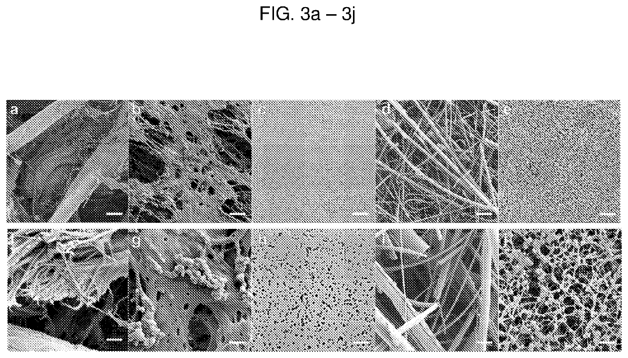
[0124]Paper membranes were chosen for analysis based on their use in either cell filtration, nucleic acid capture, or current point-of-care diagnostics. Paper samples used in this study were 3MM Chr cellulose (CHR), 0.22 μm polyethersulfone (PES), 0.2 μm track etched polycarbonate (PC), 1.6 μm binder-free glass microfiber (GF), and 0.45 μm nitrocellulose (NC). CHR, GF, and NC were purchased from GE Healthcare (Pittsburgh, Pa.), catalog numbers 3MM CHR (3030861), GF / F (1825-090), and Protran BA85 (10402506), respectively. PES and PC were purchased from Millipore EMD (Billerica, Mass.), catalog numbers GPWP04700 and GTTP14250, respectively.

[0125]SEM

[0126]Scanning electron microscopy (SEM) was performed using a ...

example 3

Device

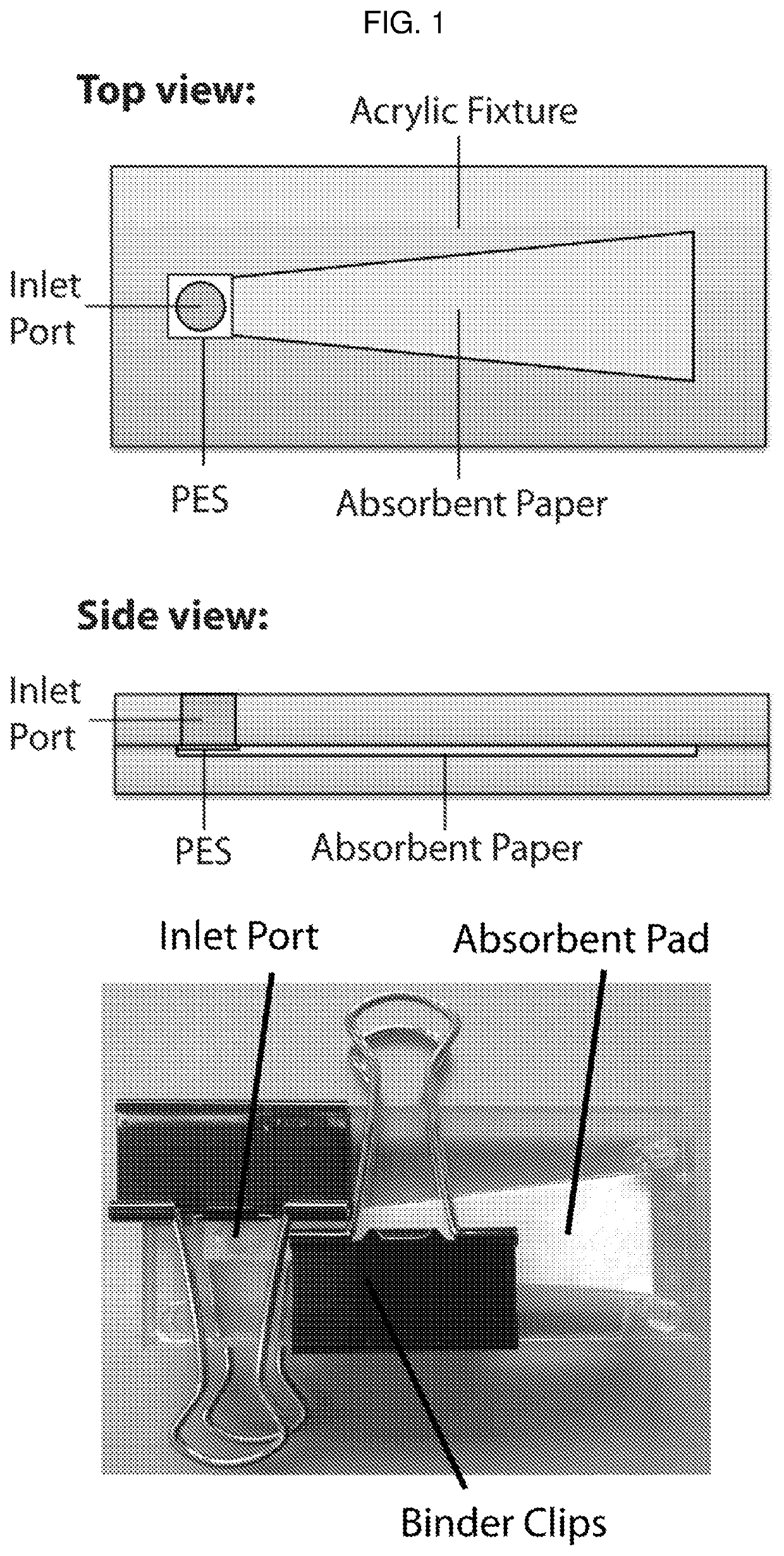
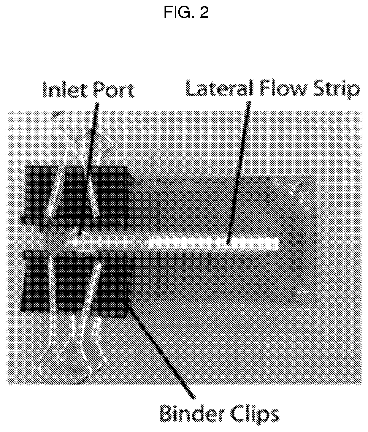
[0199]Example 3 demonstrates the construction and functionality of a device of the invention by implementing an HPV 16 DNA extraction, amplification, and detection assay directly from patient cervical samples. This on-chip HPV 16 assay addresses many of the limitations of conventional cytology by providing highly sensitive molecular level information regarding the presence of high-risk HPV 16 in cervical samples without the need for laboratory infrastructure or highly trained pathologists.

[0200]A. Materials and Methods

[0201]Human Papillomavirus 16 Cloned DNA Standards

[0202]Human Papillomavirus 16 (HPV 16) DNA standards were generated by cloning the E7 gene for HPV 16 into the pGEM-T Easy Vector (Promega, Madison, Wis.). The E7 gene was PCR amplified from HPV-16 transformed cell DNA (Advanced Biotechnologies, Inc, Eldersburg, Md.) with gene-specific forward and reverse cloning primers (Table 2) containing restriction endonuclease sequences SpeI and AatlI, respectively, using th...


PUM

PropertyMeasurementUnit
temperatureaaaaaaaaaa

Abstract

The invention features a fully-integrated rapid molecular diagnostic device that is low-cost, easy to manufacture, and simple to use. The device can serve as a molecular diagnostic platform for any disease, requiring little or no preparation or customization and can be made from simple materials (e.g., paper and adhesive film), making it inexpensive, portable, and disposable. The invention also provides methods of using the device for detection of one or more targets in a sample.

Description

STATEMENT REGARDING FEDERALLY SPONSORED RESEARCH[0001]This invention was made with Government Support under Contract Nos. AI113927, EB015403 and AI110023 awarded by the National Institutes of Health. The Government has certain rights in the invention.BACKGROUND OF INVENTION[0002]Infectious diseases disproportionately affect the developing world where standard molecular diagnostic approaches requiring expensive equipment, highly skilled technicians, and established laboratory and sample transportation infrastructure are unavailable. For example, cervical cancer is highly preventable and easily treated upon early detection, but over a half million new cases and over a quarter million deaths occur each year (Ferlay et al, Int. J. Cancer, 2010, 127:2893-2917). From a total of 528,000 new cases worldwide in 2012, 445,000 cases (84%) occurred in the developing world (Ferlay et al, Int. J. Cancer, 2014, 136, E359-E386), where, according to the World Health Organization (WHO), less than 5% ...

Claims


Application Information

Patent Timeline
Patent Type & Authority Patents(United States)
IPC IPC(8): C12Q1/6844G01N33/558B01L3/00
CPCC12Q1/6844B01L3/5023G01N33/558B01L2300/0816B01L2300/0825G01N33/54388
Inventor KLAPPERICH, CATHERINE M.RODRIGUEZ, NATALIA M.LINNES, JACQUELINE C.
Owner TRUSTEES OF BOSTON UNIV
Why Eureka
Try Eureka