Antigen responsive antibody-fluorescent dye conjugate and method for fluorescence detection and imaging of target cell using the same

a fluorescence imaging and antibody technology, applied in the direction of immunoglobulins, instruments, peptides, etc., can solve the problems of inability to distinguish whether the conjugate is bound to the target cell, fluorescent image, loss of cells, etc., and achieve the effect of dramatic enhancement of surgery accuracy and cancer treatment efficiency

Active Publication Date: 2022-01-04
NAT CANCER CENT
View PDF15 Cites 0 Cited by
  • Summary
  • Abstract
  • Description
  • Claims
  • Application Information

AI Technical Summary

Benefits of technology

[0007]An object of the present invention is to provide an antigen responsive antibody-fluorescent dye conjugate which has high binding specificity to a specific antigen present on a cancer cell surface and in which fluorescence that has been quenched, that is, a quenching effect disappears immediately upon binding to a target antigen present on the cell surface and a strong fluorescent signal is emitted, so that a cancer cell can be imaged and detected through fluorescence imaging within a short time without a washing process.
[0026]The antigen responsive antibody-fluorescent dye conjugate according to the present invention is configured such that fluorescence emittance is suppressed in a case of being present in an extracellular region, and a quenching phenomenon disappears immediately upon binding to a targeted antigen and a strong fluorescent signal can be emitted. Thus, it is possible to perform detection and imaging diagnosis of a targeted cancer cell in in vivo and ex vivo tests in a very effective manner. Therefore, in a study or screening using cancer cells, it is possible to image and detect targeted cancer cells in real-time within a short time without performing a washing process. Even in a fluorescent image-guided cancer surgery, it is contemplated that locations of cancer cells or cancer tissues are detected with high contrast within a few hours to 24 hours after intravenous administration of the antibody-fluorescent dye conjugate, so that accuracy of the surgery and therapeutic efficiency for cancer can be dramatically enhanced.

Problems solved by technology

However, for the antibody-fluorescent dye conjugates in the related art, both case of being bound to target cells and case of being near the target cells emit a strong fluorescent signal, and thus it is not possible to distinguish whether the conjugates are bound to the target cells.
That is, in an in vitro cell experiment for detecting a fluorescent image of a specific cell, after a cell or a living body tissue is treated with an antibody-fluorescent dye conjugate, a washing process is necessarily performed to remove a conjugate which is not bound to a target cell, and a fluorescent image is obtained, which is troublesome.
In particular, the washing process acts as a major limitation for a case where due to a small amount of cells to be analyzed, loss of cells with washing is concerned, a case where an analysis time is increased due to the washing process in a high-throughput drug screening, a case of suspension cells, or a case of intending to image an interaction between an antibody and a cell in real-time.
In addition, in a case of diagnosing cancer or bacteria using a microfluidics system, it is often difficult to perform a washing process.
A characteristic that both an antibody-fluorescent dye conjugate which is bound to a target cell and the antibody-fluorescent dye conjugate which is not bound to the target cell emit a strong fluorescent signal becomes more problematic in a case where the antibody-fluorescent dye is intravenously administered to a patient in order to diagnose a location of cancer with fluorescence imaging during surgery.
The antibody-fluorescent dye conjugate having a large molecular weight has a long blood half-life of a few days to a few weeks, and thus keeps a background noise signal high while circulating in blood for a long time.
Nevertheless, due to being not possible to artificially remove the antibody-fluorescent dye conjugate present in blood, there is a problem that a fluorescent image having a high tumor-to-background ratio can be obtained only in a case where the fluorescent image is obtained after waiting a few days to a few weeks until antibody-fluorescent dye conjugates present in normal tissues or bloodstream are removed sufficiently.
Thus, it takes a long time during such processes (that is, binding→intracellular migration→lysosomal degradation).
Thus, there is a disadvantage that a fluorescent image has to be obtained after waiting at least one day following administration of the target cell-specific antibody-fluorescent dye conjugate.
Furthermore, among antigens present on cell surfaces, in a case of antigens that do not allow endocytosis into cells, an antibody conjugate cannot migrate to a lysosome.
Thus, there is a problem that the target cell-specific antibody-fluorescent dye conjugate cannot be used for fluorescence imaging diagnosis of such antigens.
In addition, due to this disadvantage, the target cell-specific antibody-fluorescent dye conjugate is not suitable for obtaining an image with high contrast in an ex-vivo or in vitro cell experiment.

Method used


Image

  • Antigen responsive antibody-fluorescent dye conjugate and method for fluorescence detection and imaging of target cell using the same
  • Antigen responsive antibody-fluorescent dye conjugate and method for fluorescence detection and imaging of target cell using the same
  • Antigen responsive antibody-fluorescent dye conjugate and method for fluorescence detection and imaging of target cell using the same

Examples


example 1

[0082]1.1. Synthesis of Trastuzumab-Fluorescent Dye Conjugate

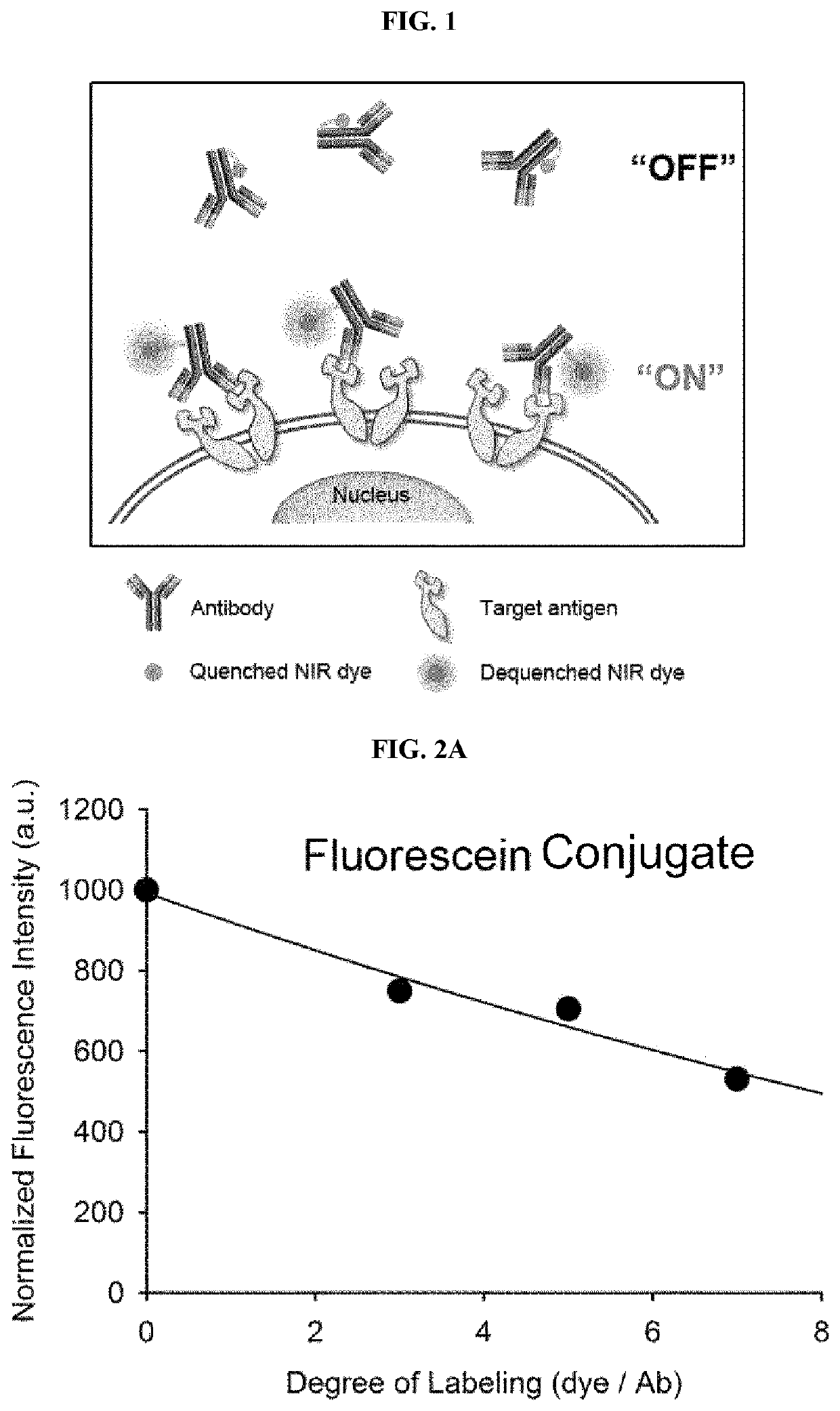
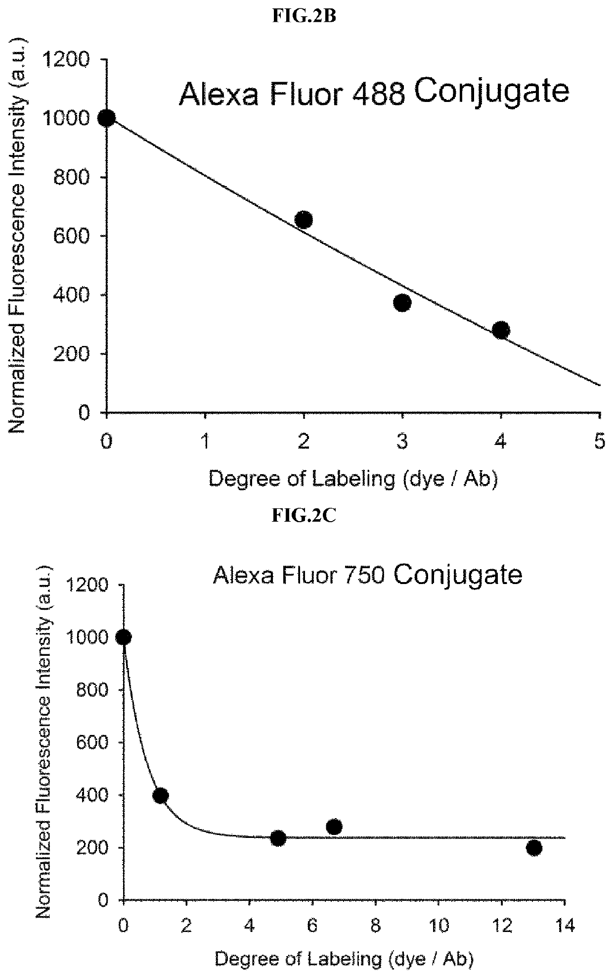
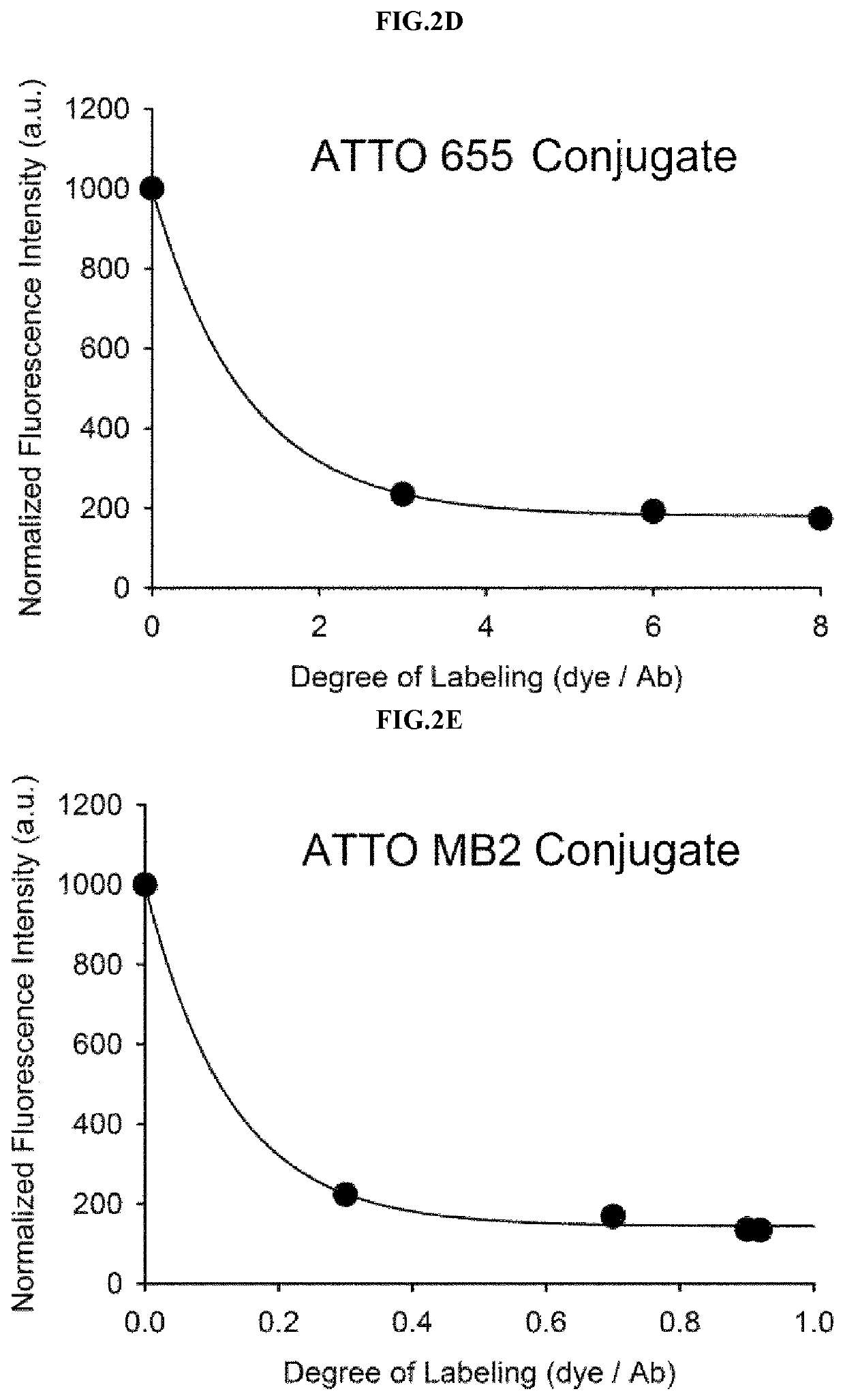
[0083]Example 1 was intended to demonstrate a basic concept and utility of an antigen responsive antibody-fluorescent dye conjugate, by using an antibody that specifically binds to Human Epidermal Growth Factor Receptor 2 (HER2) which is known to be over-expressed on surfaces of cancer cells, and causing various fluorescent dyes to be bound thereto. In particular, various fluorescent dyes are caused to be bound to Trastuzumab (Herceptin), which is a representative antibody used in clinical practice among antibodies that specifically bind to HER2, and a fluorescence-quenching effect was analyzed. For synthesis of antibody-fluorescent dye conjugates, Trastuzumab (0.5 mg, 3.4 nmol), and each of amine-reactive fluorescent dyes NHS-Fluorescein, Alexa Fluor®488 NHS ester, Alexa Fluor®750-NHS ester, ATTO 655-NHS ester, ATTO 680-NHSO ester, ATTO 700-NHS ester, a methylene blue derivative ATTO MB2-NHS ester, Indocyanine green sulfo...

example 2

[0113]2.1. Synthesis of Cetuximab-ATTO 680 Conjugate

[0114]In Example 2, in order to further demonstrate that the basic concept and utility of the antigen responsive-type antibody-fluorescent dye conjugate can be applied to various antibodies, as an example of another antibody, Cetuximab (Erbitux, manufactured by Merck Serono) which is an antibody targeting epidermal growth factor receptor (EGFR) was used to carry out a second embodiment.

[0115]It has already been shown in Example 1 that a quenching concept is realized for various fluorescent dyes. Thus, present Example 2 was intended to further verify characteristics and utility of the antigen responsive antibody-fluorescent dye conjugate, by using only ATTO 680 N-hydroxysuccinimidyl ester (ATTO 680-NHS ester) as a fluorescent dye and causing the Cetuximab and the ATTO 680 to react with each other at various ratios. For synthesis of the conjugate, the Cetuximab and the ATTO 680-NHS ester were mixed at a molar ratio of 1:1 or more and...

example 3

[0126]3.1. Synthesis of Anti-VEGF-ATTO 680 Conjugate

[0127]In Example 3, in order to further demonstrate that the basic concept and utility of the antigen responsive-type antibody-fluorescent dye conjugate can be applied to various antibodies, as an example of another antibody, anti-VEGF (ab46154, manufactured by Abcam) which targets vascular endothelial growth factor (VEGF) was used to carry out a third embodiment.

[0128]It has already been shown in Example 1 that a quenching concept is realized for various fluorescent dyes. Thus, present Example 3 was intended to further verify characteristics and utility of the antigen responsive antibody-fluorescent dye conjugate, by using only ATTO 680 N-hydroxysuccinimidyl ester (ATTO 680-NHS ester) as a fluorescent dye and causing the VEGF antibody and the ATTO 680 to react with each other at various ratios. For synthesis of the conjugate, the VEGF antibody and the ATTO 680-NHS ester were mixed at a molar ratio of 1:1 or more and dissolved in p...


PUM

PropertyMeasurementUnit
timeaaaaaaaaaa
concentrationaaaaaaaaaa

Abstract

The present invention relates to an antibody-fluorescent dye conjugate capable of cancer cell-specific fluorescence imaging diagnosis. The fluorescent dye comprises a covalently labeled antibody and is quenched by interaction with an amino acid residue such as tryptophan, tyrosine, histidine, and methionine in the antibody and upon binding of the antibody to an antigen present on a cell surface to emit fluorescence, whereby cells having a target antigen thereon can be imaged for diagnosis. When using the antibody-fluorescent dye conjugate according to the present invention during in vitro cell assays, high-throughput screening of cells, and cytodiagnosis based on microfluidics, the presence of cancer cells having a specific antigen expressed thereon can be detected at high specificity and sensitivity without a washing process, and the position of primary and metastatic cancer cells can be detected at high contrast within a short time.

Description

CROSS-REFERENCE TO RELATED APPLICATIONS[0001]This application is a continuation of International Application No. PCT / KR2017 / 002924 filed on Mar. 20, 2017, which claims priority to Korean Application No. 10-2016-0037411 filed on Mar. 29, 2016. The applications are incorporated herein by reference.TECHNICAL FIELD[0002]The present invention relates to production of an antigen responsive antibody-fluorescent dye conjugate and a target cell-specific fluorescence imaging method using the same.BACKGROUND ART[0003]Due to a great advantage that real-time image acquisition is possible while having high sensitivity as compared with magnetic resonance imaging, nuclear medicine imaging, and ultrasound imaging, a fluorescent imaging technique is evaluated as the most powerful tool which is capable of identifying, in real-time, a location of a lesion such as cancer and removing the lesion during surgery. Furthermore, as systems capable of realizing a near-infrared fluorescence imaging function in ...

Claims


Application Information

Patent Timeline
Patent Type & Authority Patents(United States)
IPC IPC(8): A61K49/00G01N33/58C07K16/18C07K16/28G01N33/574C07K16/22C07K16/30C07K16/32
CPCA61K49/0058A61K49/0028A61K49/0039A61K49/0043C07K16/18C07K16/22C07K16/2863C07K16/2878C07K16/2884C07K16/2887C07K16/3069C07K16/3092C07K16/32G01N33/57492G01N33/582C07K2317/77A61K49/0019A61K49/06G01N33/574
Inventor CHOI, YONG-DOOKIM, HYUN-JIN
Owner NAT CANCER CENT
Why Eureka
Try Eureka